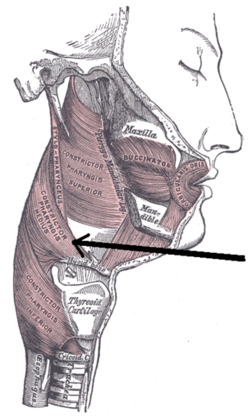
Middle pharyngeal constrictor
| Middle pharyngeal constrictor muscle | |
|---|---|

|
|

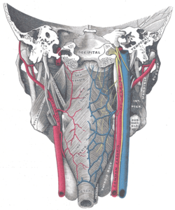
Muscles of the pharynx, viewed from behind, together with the associated vessels and nerves (middle pharyngeal constrictor muscle labeled as Mid. constr. at center)
|
|
| Details | |
| Origin | Hyoid bone |
| Insertion | Pharyngeal raphe |
| Artery | Ascending pharyngeal artery |
| Nerve | Pharyngeal plexus of vagus nerve |
| Actions | Swallowing |
| Identifiers | |
| Latin | Musculus constrictor pharyngis medius |
| TA | A05.3.01.108 |
| FMA | 46622 |
|
Anatomical terms of muscle
[]
|
|
The middle pharyngeal constrictor is a fanshaped muscle located in the neck. It is one of three pharyngeal constrictors. Similarly to the superior and inferior pharyngeal constrictor muscles, the middle pharyngeal constrictor is innervated by a branch of the vagus nerve through the pharyngeal plexus. The middle pharyngeal constrictor is smaller than the inferior pharyngeal constrictor muscle.
The middle pharyngeal constrictor arises from the whole length of the upper border of the greater cornu of the hyoid bone, from the lesser cornu, and from the stylohyoid ligament.
The fibers diverge from their origin: the lower ones descend beneath the constrictor inferior, the middle fibers pass transversely, and the upper fibers ascend and overlap the constrictor superior.
It is inserted into the posterior median fibrous raphe, blending in the middle line with the muscle of the opposite side.
As soon as the bolus of food is received in the pharynx, the elevator muscles relax, the pharynx descends, and the constrictors contract upon the bolus, and convey it downward into the esophagus. They also have respiratory mechanical effects.
Hyoid bone. Anterior surface. Enlarged.
Muscles of the neck. Lateral view.
Middle pharyngeal constrictor muscle
Middle pharyngeal constrictor muscle
Deep dissection of larynx, pharynx and tongue seen from behind
Deep dissection of larynx, pharynx and tongue seen from behind
...
Wikipedia
