Lesser cornu
| Hyoid bone | |
|---|---|

Hyoid bone — anterior surface, enlarged
|
|

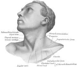
Anterolateral view of head and neck
|
|
| Details | |
| Precursor | 2nd and 3rd branchial arch |
| Identifiers | |
| Latin | os hyoideum |
| MeSH | A02.835.232.409 |
| TA | A02.1.16.001 |
| FMA | 52749 |
|
Anatomical terms of bone
[]
|
|
The hyoid bone (lingual bone or tongue-bone) (/ˈhaɪɔɪd/; Latin os hyoideum) is a horseshoe-shaped bone situated in the anterior midline of the neck between the chin and the thyroid cartilage. At rest, it lies at the level of the base of the mandible in the front and the third cervical vertebra (C3) behind.
Unlike other bones, the hyoid is only distantly articulated to other bones by muscles or ligaments. The hyoid is anchored by muscles from the anterior, posterior and inferior directions, and aids in tongue movement and swallowing. The hyoid bone provides attachment to the muscles of the floor of the mouth and the tongue above, the larynx below, and the epiglottis and pharynx behind.
Its name is derived from Greek hyoeides, meaning "shaped like the letter upsilon (υ)".
The hyoid bone is classed as an irregular bone and consists of a central part called the body, and two pairs of horns, the greater and lesser horns (or cornua).
The greater and lesser horns (Latin: cornua) are two sections of bone that project from each side of the hyoid.
The greater horns project backward from the outer borders of the body; they are flattened from above downward and taper to their end, which is a bony tubercle connecting to the lateral thyrohyoid ligament. The upper surface of the greater horns are rough and close to its lateral border, and facilitates muscular attachment. The largest of muscles that attach to the upper surface of the greater horns are the hyoglossus and the middle pharyngeal constrictor, which extend along the whole length of the horns; the digastric muscle and stylohyoid muscle have small insertions in front of these near the junction of the body with the cornu. To the medial border the thyrohyoid membrane is attached, while the anterior half of the lateral border gives insertion to the thyrohyoid muscle. The greater cornua derive from the third pharyngeal arch.
...
Wikipedia
