Mandibula
| Mandible | |
|---|---|

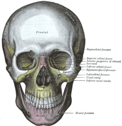
The skull from the front, with mandible shown in purple at bottom.
|
|
| Details | |
| Precursor | 1st branchial arch |
| Identifiers | |
| Latin | mandibula |
| MeSH | Mandible |
| FMA | 52748 |
|
Anatomical terms of bone
[]
|
|
The mandible,lower jaw or jawbone (from Latin mandibula, "jawbone") is the largest, strongest and lowest bone in the face. It forms the lower jaw and holds the lower teeth in place. In the midline on the anterior surface of the mandible is a faint ridge, an indication of the mandibular symphysis, where the bone is formed by the fusion of right and left processes during mandibular development. Like other symphyses in the body, this is a midline articulation where the bones are joined by fibrocartilage, but this articulation fuses together in early childhood.
The mandible consists of:
The mandible articulates with the two temporal bones at the temporomandibular joints.
Inferior alveolar nerve, branch of the mandibular division of Trigeminal (V) nerve, enters the mandibular foramen and runs forward in the mandibular canal, supplying sensation to the teeth. At the mental foramen the nerve divides into two terminal branches: incisive and mental nerves. The incisive nerve runs forward in the mandible and supplies the anterior teeth. The mental nerve exits the mental foramen and supplies sensation to the lower lip.
Rarely, a bifid inferior alveolar nerve may be present, in which case a second mandibular foramen, more inferiorly placed, exists and can be detected by noting a doubled mandibular canal on a radiograph.
The ossification of the mandible refers to the Human mandible laying down new bone material in the fibrous membrane covering the outer surfaces of Meckel's cartilages.
These cartilages form the cartilaginous bar of the mandibular arch, and are two in number, a right and a left. Their proximal or cranial ends are connected with the ear capsules, and their distal extremities are joined to one another at the symphysis by mesodermal tissue. They run forward immediately below the condyles and then, bending downward, lie in a groove near the lower border of the bone; in front of the canine tooth they incline upward to the symphysis. From the proximal end of each cartilage the malleus and incus, two of the bones of the middle ear, are developed; the next succeeding portion, as far as the lingula, is replaced by fibrous tissue, which persists to form the sphenomandibular ligament.
...
Wikipedia
