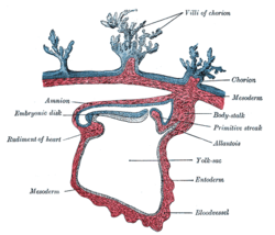
Mesodermal
| Mesoderm | |
|---|---|

Tissues derived from mesoderm.
|
|

Section through the embryo
|
|
| Details | |
| Days | 16 |
| Identifiers | |
| MeSH | A16.254.425.660 |
| FMA | 69072 |
|
Anatomical terminology
[]
|
|
In all bilaterian animals, the mesoderm is one of the three primary germ layers in the very early embryo. The other two layers are the ectoderm (outside layer) and endoderm (inside layer), with the mesoderm as the middle layer between them.
The mesoderm forms mesenchyme, mesothelium, non-epithelial blood cells and coelomocytes. Mesothelium lines coeloms. Mesoderm forms the muscles in a process known as myogenesis, septa (cross-wise partitions) and mesenteries (length-wise partitions); and forms part of the gonads (the rest being the gametes). Myogenesis is specifically a function of Mesenchyme.
The mesoderm differentiates from the rest of the embryo through intercellular signaling, after which the mesoderm is polarized by an organizing center. The position of the organizing center is in turn determined by the regions in which beta-catenin is protected from degradation by GSK-3. Beta-catenin acts as a co-factor that alters the activity of the transcription factor tcf-3 from repressing to activating, which initiates the synthesis of gene products critical for mesoderm differentiation and gastrulation. Furthermore, mesoderm has the capability to induce the growth of other structures, such as the neural plate, the precursor to the nervous system.
The mesoderm is one of the three germinal layers that appears in the third week of embryonic development. It is formed through a process called gastrulation. There are three important components, the paraxial mesoderm, the intermediate mesoderm and the lateral plate mesoderm. The paraxial mesoderm forms the somitomeres, which give rise to mesenchyme of the head and organize into somites in occipital and caudal segments. Somites give rise to the myotome (muscle tissue), sclerotome (cartilage and bone), and dermatome (subcutaneous tissue of the skin). Signals for somite differentiation are derived from surroundings structures, including the notochord, neural tube and epidermis. The intermediate mesoderm connects the paraxial mesoderm with the lateral plate, eventually it differentiates into urogenital structures consisting of the kidneys, gonads, their associated ducts, and the adrenal glands. The lateral plate mesoderm give rise to the heart, blood vessels and blood cells of the circulatory system as well as to the mesodermal component of the limbs.
...
Wikipedia
