Mammalian eye
| Eye | |
|---|---|

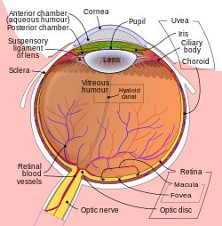
Schematic diagram of the human eye.
|
|

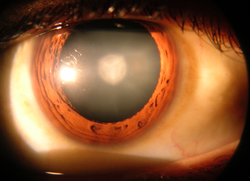
Human Eye Anterior Segment – Magnified view seen on examination with a slit lamp under diffuse illumination showing conjunctiva overlying the white sclera, transparent cornea, pharmacologically dilated pupil and cataract
|
|
| Details | |
| Latin | Oculus (plural: oculi) |
| Dorlands /Elsevier |
s_07/13264628 |
|
Anatomical terminology
[]
|
|
Mammals normally have a pair of eyes. Although mammalian vision is not so excellent as bird vision, it is at least dichromatic for most of mammalian species, with certain families (such as Hominidae) possessing a trichromatic color perception.
Dimensions vary only 1–2 mm among humans. The vertical axis is 24 mm; the transverse being larger. At birth it is generally 16–17 mm, enlarging to 22.5–23 mm by three years of age. Between then and age 13 the eye attains its mature size. It weighs 7.5 grams and its volume is roughly 6.5 ml. Along a line through the nodal (central) point of the eye is the optic axis, which is slightly five degrees toward the nose from the visual axis (i.e., that going towards the focused point to the fovea.
The structure of the mammalian eye can be divided into three main layers or tunics whose names reflect their basic functions: the fibrous tunic, the vascular tunic, and the nervous tunic.
The mammalian eye can also be divided into two main segments: the anterior segment and the posterior segment.
The human eye is not a plain sphere but is like two spheres combined, a smaller, more sharply curved one and a larger lesser curved sphere. The former, the anterior segment is the front sixth of the eye that includes the structures in front of the vitreous humour: the cornea, iris, ciliary body, and lens.
Within the anterior segment are two fluid-filled spaces:
Aqueous humor fills these spaces within the anterior segment and provides nutrients to the surrounding structures.
...
Wikipedia
