Lung bud
| Lung bud | |
|---|---|

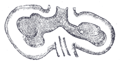
Bronchial buds from a human embryo of about four weeks, showing commencing lobulations.
|
|

Lungs of a human embryo at about six weeks
|
|
| Details | |
| Days | 28 |
| Precursor | Ventral part of the foregut |
| Identifiers | |
| Latin | Gemma respiratoria, gemma pulmonalis |
| Code | TE E5.5.3.0.0.0.3 |
|
Anatomical terminology
[]
|
|
The lung bud forms from the respiratory diverticulum (sometimes referred to as the lung bud) as an embryological endodermal structure that develops into the respiratory tract organs such as the larynx, trachea, bronchi and lungs. It arises from part of the laryngotracheal tube.
In the fourth week of development, the respiratory diverticulum, starts to grow from the ventral (front) side of the foregut into the mesoderm that surrounds it, forming the lung bud. Around the 28th day, during the separation of the lung bud from the foregut it forms the trachea and splits into two bronchial buds, one on each side.
The molecular signaling involved in the specification of the respiratory bud starts with the expression of the Nbx2-1 gene, which determines the respiratory field – the area where the respiratory bud will begin to grow from. The signaling that makes the growth of the respiratory bud possible is complex and involves a number of interactions between the mesoderm and the respiratory bud epithelium, in which members of the Fgf and Fgfr family of genes express.
At first, the posterior part of the trachea is open to the esophagus, but as the bud elongates two longitudinal mesodermal ridges known as the laryngotracheal folds, begin to form and grow until they join, forming a wall between the two organs. An incomplete separation of the organs leads to a congenital abnormality known as a tracheoesophageal fistula.
...
Wikipedia
