Luciferase
| Dinoflagellate Luciferase catalytic domain |
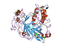
crystal structure of a luciferase domain from the dinoflagellate Lingulodinium polyedrum
|
| Identifiers |
| Symbol |
Luciferase_cat |
| Pfam |
PF10285 |
| InterPro |
IPR018804 |
|
|
| Dinoflagellate Luciferase helical bundle domain |
|
crystal structure of a Dinoflagellate luciferase domain from the dinoflagellate Lingulodinium polyedrum
|
| Identifiers |
| Symbol |
Luciferase_3H |
| Pfam |
PF10284 |
| InterPro |
IPR018475 |
|
|
Luciferase is a generic term for the class of oxidative enzymes that produce bioluminescence, and is distinct from a photoprotein. The name is derived from the Latin word lucifer (lightbringer). One example is the firefly luciferase (EC 1.13.12.7) from the firefly Photinus pyralis. "Firefly luciferase" as a laboratory reagent often refers to P. pyralis luciferase although recombinant luciferases from several other species of fireflies are also commercially available.
A variety of organisms regulate their light production using different luciferases in a variety of light-emitting reactions. The most famous are the fireflies, although the enzyme exists in organisms as different as the Jack-O-Lantern mushroom (Omphalotus olearius) and many marine creatures.
The luciferases of fireflies - of which there are over 2000 species - and of the other Elateroidea (click beetles and relatives in general) are diverse enough to be useful in molecular phylogeny. In fireflies, the oxygen required is supplied through a tube in the abdomen called the abdominal trachea. One well-studied luciferase is that of the Photinini firefly Photinus pyralis, which has an optimum pH of 7.8.
Also well studied is the fancy sea pansy, Renilla reniformis. In this organism, the luciferase (Renilla-luciferin 2-monooxygenase) is closely associated with a luciferin-binding protein as well as a green fluorescent protein (GFP). Calcium triggers release of the luciferin (coelenterazine) from the luciferin binding protein. The substrate is then available for oxidation by the luciferase, where it is degraded to coelenteramide with a resultant release of energy. In the absence of GFP, this energy would be released as a photon of blue light (peak emission wavelength 482 nm). However, due to the closely associated GFP, the energy released by the luciferase is instead coupled through resonance energy transfer to the fluorophore of the GFP, and is subsequently released as a photon of green light (peak emission wavelength 510 nm). The catalyzed reaction is:
...
Wikipedia


