Intima
| Tunica intima | |
|---|---|
 |
|

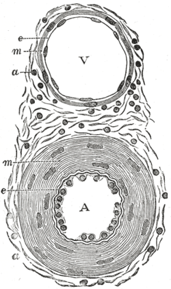
Transverse section through a small artery and vein of the mucous membrane of the epiglottis of a child. (Tunica intima is at "e")
|
|
| Details | |
| Identifiers | |
| Latin | Tunica intima |
| MeSH | A07.231.330.800 |
| Code | TH H3.09.02.0.01003 |
| TA | A12.0.00.018 |
| FMA | 55589 |
|
Anatomical terminology
[]
|
|
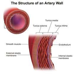
The tunica intima (New Latin "inner coat"), or intima for short, is the innermost tunica (layer) of an artery or vein. It is made up of one layer of endothelial cells and is supported by an internal elastic lamina. The endothelial cells are in direct contact with the blood flow.
The tunicae of blood vessels are three layers—an inner layer (the tunica intima), a middle layer (the tunica media), and an outer layer (the tunica adventitia).
In dissection, the inner coat (tunica intima) can be separated from the middle (tunica media) by a little , or it may be stripped off in small pieces; but, because of its friability, it cannot be separated as a complete membrane. It is a fine, transparent, colorless structure which is highly elastic, and, after death, is commonly corrugated into longitudinal wrinkles.
The inner coat consists of:
Vein
Microphotography of arterial wall with calcified (violet colour) atherosclerotic plaque (haematoxillin and eosin stain)
This article incorporates text in the public domain from the 20th edition of Gray's Anatomy (1918)
...
Wikipedia
