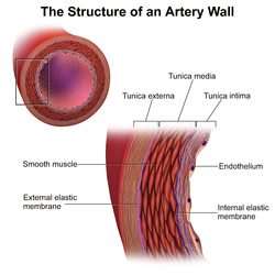
Tunica media
| Tunica media | |
|---|---|
 |
|

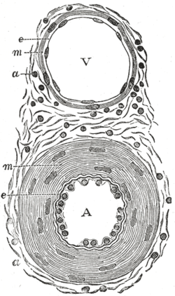
Transverse section through a small artery and vein of the mucous membrane of the epiglottis of a child. (Tunica media is at 'm')
|
|
| Details | |
| Identifiers | |
| Latin | tunica media vasorum |
| MeSH | A02.633.570.491.800 |
| Code | TH H3.09.02.0.01007 |
| Dorlands /Elsevier |
t_22/12831907 |
| TA | A12.0.00.019 |
| FMA | 55590 |
|
Anatomical terminology
[]
|
|
The tunica media (New Latin "middle coat"), or media for short, is the middle tunica (layer) of an artery or vein. It lies between the tunica intima on the inside and the tunica externa on the outside.
Tunica media is made up of smooth muscle cells and elastic tissue. It lies between the tunica intima on the inside and the tunica externa on the outside.
The middle coat (tunica media) is distinguished from the inner (tunica intima) by its color and by the transverse arrangement of its fibers.
The middle coat is composed of a thick layer of connective tissue with elastic fibers, intermixed, in some veins, with a transverse layer of muscular tissue.
The white fibrous element is in considerable excess, and the elastic fibers are in much smaller proportion in the veins than in the arteries.
This article incorporates text in the public domain from the 20th edition of Gray's Anatomy (1918)
Artery wall
Vein
Section of a medium-sized artery
Microphotography of arterial wall with calcified (violet colour) atherosclerotic plaque (haematoxillin & eosin stain)
...
Wikipedia
