Internal auditory canal
| Internal auditory meatus | |
|---|---|

Left temporal bone. Inner surface.
|
|

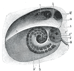
Diagrammatic view of the fundus of the right internal acoustic meatus.
|
|
| Details | |
| Identifiers | |
| Latin | meatus acusticus internus |
| TA | A02.1.06.033 |
| FMA | 53163 |
|
Anatomical terms of bone
[]
|
|
The internal auditory meatus (also meatus acusticus internus, internal acoustic meatus, internal auditory canal, or internal acoustic canal) is a canal within the petrous part of the temporal bone of the skull between the posterior cranial fossa and the inner ear.
The internal auditory meatus provides a passage through which the vestibulocochlear nerve, the facial nerve, and the labyrinthine artery (an internal auditory branch of the basilar artery) can pass from inside the skull to structures of the inner ear and face.
It also contains the vestibular ganglion.
The opening to the meatus is called the porus acusticus internus, or its English translation, the internal acoustic opening. It is located inside the posterior cranial fossa of the skull, near the center of the posterior surface of the petrous part of the temporal bone. The size varies considerably; its margins are smooth and rounded.
The internal auditory meatus is short (about 1 cm) and runs laterally into the bone.
At its end are the openings for three different canals, one of which is the facial canal. The facial nerve travels through the facial canal, eventually exiting the skull at the stylomastoid foramen.
The antero-superior part transmits the facial nerve and nervus intermedius and is separated from the postero-superior section, which transmits the superior vestibular nerve, by Bill's bar (named by William F. House). The falciform crest or transverse crest separates the superior part from the inferior part. The cochlear nerve runs antero-inferiorly and the inferior vestibular nerve runs postero-inferiorly.
...
Wikipedia
