Inner ear
| Inner ear | |
|---|---|
 |
|
| Details | |
| Artery | labyrinthine artery |
| Identifiers | |
| Latin | auris interna |
| MeSH | A09.246.631 |
| TA | A15.3.03.001 |
| FMA | 60909 |
|
Anatomical terminology
[]
|
|
| Organ of Corti | |
|---|---|

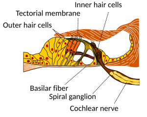
A cross section of the cochlea illustrating the organ of Corti.
|
|

Section through the spiral organ of Corti. Magnified.
|
|
| Identifiers | |
| TA | A15.3.03.001 |
| FMA | 60909 |
|
Anatomical terminology
[]
|
|
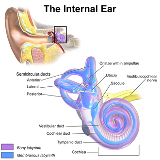
The inner ear (internal ear, auris interna) is the innermost part of the vertebrate ear. In vertebrates, the inner ear is mainly responsible for sound detection and balance. In mammals, it consists of the bony labyrinth, a hollow cavity in the temporal bone of the skull with a system of passages comprising two main functional parts:
The inner ear is found in all vertebrates, with substantial variations in form and function. The inner ear is innervated by the eighth cranial nerve in all vertebrates.
The labyrinth can be divided by layer or by region.
The bony labyrinth, or osseous labyrinth, is the network of passages with bony walls lined with periosteum. The membranous labyrinth runs inside of the bony labyrinth. There is a layer of perilymph fluid between them. The three parts of the bony labyrinth are the vestibule of the ear, the semicircular canals, and the cochlea.
In the middle ear, the energy of pressure waves is translated into mechanical vibrations by the three auditory ossicles. Pressure waves move the tympanic membrane which in turns moves the malleus, the first bone of the middle ear. The malleus articulates to incus which connects to the stapes. The footplate of the stapes connects to the oval window, the beginning of the inner ear. When the stapes presses on the oval window, it causes the perilymph, the liquid of the inner ear to move. The middle ear thus serves to convert the energy from sound pressure waves to a force upon the perilymph of the inner ear. The oval window has only approximately 1/18 the area of the tympanic membrane and thus produces a higher pressure. The cochlea propagates these mechanical signals as waves in the fluid and membranes, and then converts them to nerve impulses which are transmitted to the brain.
The vestibular system is the region of the inner ear where the semicircular canals converge, close to the cochlea. The vestibular system works with the visual system to keep objects in view when the head is moved. Joint and muscle receptors are also important in maintaining balance. The brain receives, interprets, and processes the information from all these systems to create the sensation of balance.
...
Wikipedia
