Herpes encephalitis
| Herpesviral encephalitis | |
|---|---|
 |
|
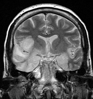
| coronal T2-weighted MR image shows high signal in the temporal lobes including hippocampal formations and parahippogampal gyrae, insulae, and right inferior frontal gyrus. A brain biopsy was performed and the histology was consistent with encephalitis. PCR was repeated on the biopsy specimen and was positive for HSV | |
| Classification and external resources | |
| ICD-10 | B00.4 |
| ICD-9-CM | 054.3 |
| eMedicine | article/1165183 article/341142 |
Herpesviral encephalitis is encephalitis due to herpes simplex virus.
Herpes simplex encephalitis (HSE) is a viral infection of the human central nervous system. It is estimated to affect at least 1 in 500,000 individuals per year and some studies suggest an incidence rate of 5.9 cases per 100,000 live births. The majority of cases of herpes encephalitis are caused by herpes simplex virus-1 (HSV-1), the same virus that causes cold sores. 57% of American adults are infected with HSV-1, which is spread through droplets, casual contact, and sometimes sexual contact, though most infected people never have cold sores. About 10% of cases of herpes encephalitis are due to HSV-2, which is typically spread through sexual contact. About 1 in 3 cases of HSE result from primary HSV-1 infection, predominantly occurring in individuals under the age of 18; 2 in 3 cases occur in seropositive persons, few of whom have history of recurrent orofacial herpes. Approximately 50% of individuals who develop HSE are over 50 years of age.
Most individuals with HSE show a decrease in their level of consciousness and an altered mental state presenting as confusion, and changes in personality. Increased numbers of white blood cells can be found in patient's cerebrospinal fluid, without the presence of pathogenic bacteria and fungi. Patients typically have a fever and may have seizures. The electrical activity of the brain changes as the disease progresses, first showing abnormalities in one temporal lobe of the brain, which spread to the other temporal lobe 7–10 days later. Imaging by CT or MRI shows characteristic changes in the temporal lobes (see Figure). Definite diagnosis requires testing of the cerebrospinal fluid (CSF) by a lumbar puncture (spinal tap) for presence of the virus. The testing takes several days to perform, and patients with suspected Herpes encephalitis should be treated with acyclovir immediately while waiting for test results.
HSE is thought to be caused by the transmission of virus from a peripheral site on the face following HSV-1 reactivation, along a nerve axon, to the brain. The virus lies dormant in the ganglion of the trigeminal cranial nerve, but the reason for reactivation, and its pathway to gain access to the brain, remains unclear, though changes in the immune system caused by stress clearly play a role in animal models of the disease. The olfactory nerve may also be involved in HSE, which may explain its predilection for the temporal lobes of the brain, as the olfactory nerve sends branches there. In horses, a single-nucleotide polymorphism is sufficient to allow the virus to cause neurological disease; but no similar mechanism has been found in humans.
...
Wikipedia
