Hereditary spherocytosis
| Hereditary spherocytosis | |
|---|---|
 |
|
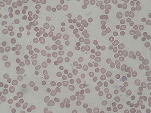
| Peripheral blood smear from patient with hereditary spherocytosis | |
| Classification and external resources | |
| Specialty | hematology |
| ICD-10 | D58.0 |
| ICD-9-CM | 282.0 |
| OMIM | 182900 |
| DiseasesDB | 5827 |
| MedlinePlus | 000530 |
| eMedicine | med/2147 |
| MeSH | D013103 |
Hereditary spherocytosis (also known as Minkowski–Chauffard syndrome) is an autosomal dominant abnormality of erythrocytes. The disorder is caused by mutations in genes relating to membrane proteins that allow for the erythrocytes to change shape. The abnormal erythrocytes are sphere-shaped (spherocytosis) rather than the normal biconcave disk shaped. Dysfunctional membrane proteins interfere with the cell's ability to be flexible to travel from the arteries to the smaller capillaries. This difference in shape also makes the red blood cells more prone to rupture. Cells with these dysfunctional proteins are taken for degradation at the spleen. This shortage of erythrocytes results in hemolytic anemia.
It was first described in 1871 and is the most common cause of inherited hemolysis in Europe and North America within the Caucasian population, with an incidence of 1 in 5000 births. The clinical severity of HS varies from symptom-free carrier to severe haemolysis because the disorder exhibits incomplete penetrance in its expression.
Symptoms include anemia, jaundice, splenomegaly, and fatigue. On a blood smear, Howell-Jolly bodies may be seen within red blood cells. Primary treatment for patients with symptomatic HS has been total splenectomy, which eliminates the hemolytic process, allowing normal hemoglobin, reticulocyte and bilirubin levels.
As in non-hereditary spherocytosis, the spleen destroys the spherocytes. This process of red blood cells rupturing directly results in varying degrees of anemia (causing a pale appearance and fatigue), high levels of bilirubin in the blood (causing jaundice), and splenomegaly.
...
Wikipedia
