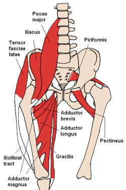
Gracilis muscle
| Gracilis muscle | |
|---|---|

The gracilis and nearby muscles
|
|

Gracilis labeled at far right.
|
|
| Details | |
| Origin | ischiopubic ramus |
| Insertion | tibia (pes anserinus) |
| Artery | medial circumflex femoral artery |
| Nerve | anterior branch of obturator nerve |
| Actions | flexes, medially rotates, and adducts the hip, |
| Identifiers | |
| Latin | musculus gracilis |
| Dorlands /Elsevier |
m_22/12549236 |
| TA | A04.7.02.030 |
| FMA | 43882 |
|
Anatomical terms of muscle
[]
|
|
The gracilis (/ˈɡræsᵻlᵻs/) (Latin for "slender") is the most superficial muscle on the medial side of the thigh. It is thin and flattened, broad above, narrow and tapering below.
It arises by a thin aponeurosis from the anterior margins of the lower half of the symphysis pubis and the upper half of the pubic arch.
The muscle's fibers run vertically downward, ending in a rounded tendon. This tendon passes behind the medial condyle of the femur, curves around the medial condyle of the tibia where it becomes flattened, and inserts into the upper part of the medial surface of the body of the tibia, below the condyle. For this reason, the muscle is a lower limb adductor. At its insertion the tendon is situated immediately above that of the semitendinosus muscle, and its upper edge is overlapped by the tendon of the sartorius muscle, which it joins to form the pes anserinus. The pes anserinus is separated from the medial collateral ligament of the knee-joint by a bursa.
A few of the fibers of the lower part of the tendon are prolonged into the deep fascia of the leg.
By its inner or superficial surface gracilis is in relation with the fascia lata, and below with the sartorius and internal saphenous nerve; the internal saphenous vein crosses it lying superficially to the fascia lata.
...
Wikipedia
