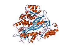
Glutathione synthetase
Glutathione synthetase (GSS) (EC 6.3.2.3) is the second enzyme in the glutathione (GSH) biosynthesis pathway. It catalyses the condensation of gamma-glutamylcysteine and glycine, to form glutathione. Glutathione synthetase is also a potent antioxidant. It is found in a large number of species including bacteria, yeast, mammals, and plants.
In humans, defects in GSS are inherited in an autosomal recessive way and are the cause of severe metabolic acidosis, 5-oxoprolinuria, increased rate of haemolysis, and defective function of the central nervous system. Deficiencies in GSS can cause a spectrum of deleterious symptoms in plants and human beings alike.
In eukaryotes, this is a homodimeric enzyme. The substrate-binding domain has a 3-layer alpha/beta/alpha structure. This enzyme utilizes and stabilizes an acylphosphate intermediate to later perform a favorable nucleophilic attack of glycine.
Human and yeast glutathione synthetases are homodimers, meaning they are composed of two identical subunits of itself non-covalently bound to each other. On the other hand, E. coli glutathione synthetase is a homotetramer. Nevertheless, they are part of the ATP-grasp superfamily, which consists of 21 enzymes that contain an ATP-grasp fold. Each subunit interacts with each other through alpha helix and beta sheet hydrogen bonding interactions and contains two domains. One domain facilitates the ATP-grasp mechanism and the other is the catalytic active site for γ-glutamylcysteine. The ATP-grasp fold is conserved within the ATP-grasp superfamily and is characterized by two alpha helices and beta sheets that hold onto the ATP molecule between them. The domain containing the active site exhibits interesting properties of specificity. In contrast to γ-glutamylcysteine synthetase, glutathione synthetase accepts a large variety of glutamyl-modified analogs of γ-glutamylcysteine, but is much more specific for cysteine-modified analogs of γ-glutamylcysteine. Crystalline structures have shown glutathione synthetase bound to GSH, ADP, two magnesium ions, and a sulfate ion. Two magnesium ions function to stabilize the acylphosphate intermediate, facilitate binding of ATP, and activate removal of phosphate group from ATP. Sulfate ion serves as a replacement for inorganic phosphate once the acylphosphate intermediate is formed inside the active site.
...
Wikipedia


