Globin
| Globin family | |||||||||
|---|---|---|---|---|---|---|---|---|---|

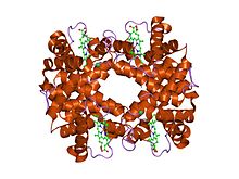
the Structure of deoxyhemoglobin Rothschild 37 beta Trp----Arg: a mutation that creates an intersubunit chloride-binding site.
|
|||||||||
| Identifiers | |||||||||
| Symbol | Globin | ||||||||
| Pfam | PF00042 | ||||||||
| Pfam clan | CL0090 | ||||||||
| InterPro | IPR000971 | ||||||||
| PROSITE | PS01033 | ||||||||
| SCOP | 1hba | ||||||||
| SUPERFAMILY | 1hba | ||||||||
| CDD | cd01067 | ||||||||
|
|||||||||
| Available protein structures: | |
|---|---|
| Pfam | structures |
| PDB | RCSB PDB; PDBe; PDBj |
| PDBsum | structure summary |
| Bacterial-like Globin | |||||||||
|---|---|---|---|---|---|---|---|---|---|

crystal structure of "truncated" hemoglobin n (hbn) from mycobacterium tuberculosis, soaked with xe atoms
|
|||||||||
| Identifiers | |||||||||
| Symbol | Bac_globin | ||||||||
| Pfam | PF01152 | ||||||||
| Pfam clan | CL0090 | ||||||||
| InterPro | IPR001486 | ||||||||
| PROSITE | PDOC00933 | ||||||||
| SCOP | 1dlw | ||||||||
| SUPERFAMILY | 1dlw | ||||||||
|
|||||||||
| Available protein structures: | |
|---|---|
| Pfam | structures |
| PDB | RCSB PDB; PDBe; PDBj |
| PDBsum | structure summary |
The globins are a superfamily of heme-containing globular proteins, involved in binding and/or transporting oxygen. These proteins all incorporate the globin fold, a series of eight alpha helical segments. Two prominent members include myoglobin and hemoglobin. Both of these proteins reversibly bind oxygen via a heme prosthetic group. They are widely distributed in many organisms.
Globin superfamily members share a common three-dimensional fold. This 'globin fold' typically consists of eight alpha helices, although some proteins have additional helix extensions at their termini. Since the globin fold contains only helices, it is classified as an all-alpha protein fold.
The globin fold is found in its namesake globin families as well as in phycocyanins. The globin fold was thus the first protein fold discovered (myoglobin was the first protein whose structure was solved).
The eight helices of the globin fold core share significant nonlocal structure, unlike other structural motifs in which amino acids close to each other in primary sequence are also close in space. The helices pack together at an average angle of about 50 degrees, significantly steeper than other helical packings such as the helix bundle. The exact angle of helix packing depends on the sequence of the protein, because packing is mediated by the sterics and hydrophobic interactions of the amino acid side chains near the helix interfaces.
...
Wikipedia
