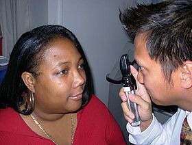
Fundoscopy
| Ophthalmoscopy | |
|---|---|
| Intervention | |

Ophthalmoscopic exam: the medical provider would next move in and observe with the ophthalmoscope from a distance of one to several cm.
|
|
| MeSH | D009887 |
Ophthalmoscopy, also called funduscopy, is a test that allows a health professional to see inside the fundus of the eye and other structures using an ophthalmoscope (or funduscope). It is done as part of an eye examination and may be done as part of a routine physical examination. It is crucial in determining the health of the retina, optic disc, and vitreous humor.
The pupil is a hole through which the eye's interior will be viewed. Opening the pupil wider (dilating it) is a simple and effective way to better see the structures behind it. Therefore, dilation of the pupil (mydriasis) is often accomplished with medicated eye drops before funduscopy. However, although dilated fundus examination is ideal, undilated examination is more convenient and is also helpful (albeit not as comprehensive), and it is the most common type in primary care.
An alternative or complement to ophthalmoscopy is to perform a fundus photography, where the image can be analysed later by a professional.
It is of two major types:
Each type of ophthalmoscopy has a special type of ophthalmoscope:
Ophthalmoscopy is done as part of a routine physical or complete eye examination.
It is used to detect and evaluate symptoms of various retinal vascular diseases or eye diseases such as glaucoma.
In patients with headaches, the finding of swollen optic discs, or papilledema, on ophthalmoscopy is a key sign, as this indicates raised intracranial pressure (ICP) which could be due to hydrocephalus, benign intracranial hypertension (aka pseudotumor cerebri) or brain tumor, amongst other conditions. Cupped optic discs are seen in glaucoma.
...
Wikipedia
