Foramen ovale (heart)
| Foramen ovale (heart) | |
|---|---|

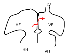
Sketch showing foramen ovale in a fetal heart. Red arrow shows blood from the inferior cava traveling to the right atrium and then to the left atrium. HF: right atrium, VF: left atrium. HH and VH: right and left ventricle. The heart still has a common pulmonary vein (LV), instead of four.
|
|

Heart of human embryo of about thirty-five days, opened on left side.
|
|
| Details | |
| Precursor | Septum secundum |
| System | Cardiovascular system |
| Identifiers | |
| Acronym(s) | PFO |
| MeSH | A07.541.459 |
| TA | A12.1.01.007 |
| FMA | 86043 |
|
Anatomical terminology
[]
|
|
In the fetal heart, the foramen ovale (/fəˈreɪmən oʊˈvæli, -mɛn-, -ˈvɑː-, -ˈveɪ-/), also foramen Botalli, ostium secundum of Born or falx septi, allows blood to enter the left atrium from the right atrium. It is one of two fetal cardiac shunts, the other being the ductus arteriosus (which allows blood that still escapes to the right ventricle to bypass the pulmonary circulation). Another similar adaptation in the fetus is the ductus venosus. In most individuals, the foramen ovale closes at birth. It later forms the fossa ovalis.
A fetus receives oxygen not from its lungs, but from the mother's oxygen-rich blood via the placenta. Oxygenated blood from the placenta travels through the umbilical cord to the right atrium of the fetal heart. As the fetal lungs are non-functional at this time, it is more efficient for the blood to bypass them. This is accomplished through 2 cardiac shunts. The first is the foramen ovale which shunts blood from the right atrium to the left atrium. The second is the ductus arteriosus which shunts blood from the pulmonary artery (which, after birth, carries blood from the right side of the heart to the lungs) to the descending aorta.
...
Wikipedia
