Atrium (heart)
| Atrium | |
|---|---|

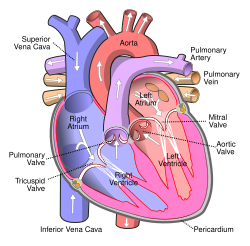
Front view of heart showing the atria
|
|
| Details | |
| Identifiers | |
| Latin | Atrium |
| TA | A12.1.00.017 |
| FMA | 7099 |
|
Anatomical terminology
[]
|
|
The atrium (plural: atria) is one of the two blood collection chambers of the heart. It was previously called the auricle, but that name is now used for an appendage of the atrium. The atrium is a chamber in which blood enters the heart, as opposed to the ventricle, where it is pushed out of the organ. It has a thin-walled structure that allows blood to return to the heart. There is at least one atrium in animals with a closed circulatory system.
The atrium receives blood as it returns to the heart to complete a circulating cycle, whereas the ventricle pumps blood out of the heart to start a new cycle.
Humans have a four-chambered heart consisting of the right atrium, left atrium, right ventricle, and left ventricle. The atria, are the two upper chambers. The right atrium receives and holds deoxygenated blood from the superior vena cava, inferior vena cava, anterior cardiac veins and smallest cardiac veins and the coronary sinus, which it then sends down to the right ventricle (through the tricuspid valve) which in turn sends it to the pulmonary artery for pulmonary circulation. The left atrium receives the oxygenated blood from the left and right pulmonary veins, which it pumps to the left ventricle (through the mitral valve) for pumping out through the aorta for systemic circulation.
The right atrium and right ventricle are often referred to as the right heart and similarly the left atrium and left ventricle are often referred to as the left heart. The atria do not have valves at their inlets and as a result, a venous pulsation is normal and can be detected in the jugular vein as the jugular venous pressure. Internally, there are the rough pectinate muscles and crista terminalis of His, which act as a boundary inside the atrium and the smooth walled part of the right atrium, the sinus venarum derived from the sinus venosus. The sinus venarum is the adult remnant of the sinus venous and it surrounds the openings of the venae cavae and the coronary sinus. Attached to the right atrium is the right atrial appendage – a pouch-like extension of the pectinate muscles. The interatrial septum separates the right atrium from the left atrium and this is marked by a depression in the right atrium –the fossa ovalis. The atria are depolarised by calcium.
...
Wikipedia
