Flocculus (cerebellar)
| Flocculus | |
|---|---|

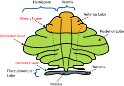
Schematic representation of the major anatomical subdivisions of the cerebellum. Superior view of an "unrolled" cerebellum, placing the vermis in one plane.
|
|

Anterior view of the cerebellum. ("Flocculus" labeled at upper right.)
|
|
| Details | |
| Part of | Cerebellum |
| System | Vestibular |
| Artery | AICA |
| Identifiers | |
| NeuroNames | hier-677 |
| NeuroLex ID | Flocculus |
| Dorlands /Elsevier |
f_09/12368554 |
| TA | A14.1.07.304 |
| FMA | 83881 |
|
Anatomical terms of neuroanatomy
[]
|
|
The flocculus (Latin: tuft of wool, diminutive) is a small lobe of the cerebellum at the posterior border of the middle cerebellar peduncle anterior to the biventer lobule. Like other parts of the cerebellum, the flocculus is involved in motor control. It is an essential part of the vestibulo-ocular reflex, and aids in the learning of basic motor skills in the brain.
It is associated with the nodulus of the vermis; together, these two structures compose the vestibular part of the cerebellum.
At its base, the flocculus receives input from the inner ear's vestibular system and regulates balance. Many floccular projections connect to the motor nuclei involved in control of eye movement.
The flocculus is contained within the flocculonodular lobe which is connected to the cerebellum. The cerebellum is the section of the brain that is essential for motor control. As a part of the cerebellum, the flocculus plays a part of the vestibulo-ocular reflex system, a system that controls the movement of the eye in coordination with movements of the head. There are five separate “zones” in the flocculus and two halves, the caudal and rostral half.
The flocculus has a complex circuitry that is reflected in the structure of the zones and halves. These "zones" of the flocculus refer to five separate groupings of Purkinje cells that project to different areas of the brain. Depending upon where stimulus occurs in the flocculus, signals can be projected to very different parts of the brain. The first and third zones of the flocculus project to the superior vestibular nucleus, the second and fourth zone projects to the medial vestibular nucleus, and the fifth zone projects to the interposed posterior nucleus, a part of the cerebellum.
The anatomy of the flocculus shows that it is composed of two disjointed lobes or halves. The “halves” of the flocculus refer to the caudal half and the rostral half, and they indicate from where fiber projections are received and the path in which a signal travels. The caudal half of the flocculus receives mossy fiber projections mainly from the vestibular system and tegmental pontine reticular nucleus, an area within the floor of the midbrain that affects the axonal projections or images received by the cerebellum. Vestibular inputs are also carried through climbing fibers that project into the flocculus, stimulating Purkinje cells. Leading research would suggest that climbing fibers play a specific role in motor learning. The climbing fibers then send the image or projection to the part of the brain that receives electrical signals and generates movement. From the midbrain, corticopontine fibers carry information from the primary motor cortex. From there, projections are sent to the ipsilateral pontine nucleus in the ventral pons, both of which are associated with projections to the cerebellum. Finally, pontocerebellar projections carry vestibulo-occular signals to the contralateral cerebellum via the middle cerebellar peduncle. The rostral half of the flocculus also receives mossy fiber projections from the pontine nuclei; however, it receives very little projection from the vestibular system.
...
Wikipedia
