Fatty liver
| Fatty liver | |
|---|---|
| Synonyms | fatty liver disease (FLD), hepatic steatosis |
 |
|
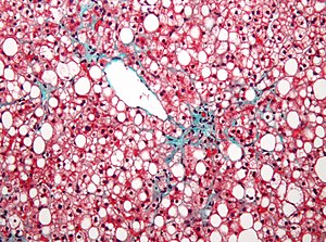
| Micrograph showing a fatty liver (macrovesicular steatosis), as seen in non-alcoholic fatty liver disease. Trichrome stain. | |
| Classification and external resources | |
| Specialty | Gastroenterology |
| ICD-10 | K70, K76.0 |
| ICD-9-CM | 571.0, 571.8 |
| DiseasesDB | 18844 |
| eMedicine | med/775 article/170409 |
| MeSH | C06.552.241 |
Fatty liver is a reversible condition wherein large vacuoles of triglyceride fat accumulate in via the process of steatosis (i.e., abnormal retention of lipids within a cell). Despite having multiple causes, fatty liver can be considered a single disease that occurs worldwide in those with excessive alcohol intake and the obese (with or without effects of insulin resistance). The condition is also associated with other diseases that influence fat metabolism. When this process of fat metabolism is disrupted, the fat can accumulate in the liver in excessive amounts, thus resulting in a fatty liver. It is difficult to distinguish alcoholic FLD from nonalcoholic FLD, and both show microvesicular and macrovesicular fatty changes at different stages.
Accumulation of fat may also be accompanied by a progressive inflammation of the liver (hepatitis), called steatohepatitis. By considering the contribution by alcohol, fatty liver may be termed alcoholic steatosis or nonalcoholic fatty liver disease (NAFLD), and the more severe forms as alcoholic steatohepatitis (part of alcoholic liver disease) and non-alcoholic steatohepatitis (NASH).
Fatty liver (FL) is commonly associated with alcohol or metabolic syndrome (diabetes, hypertension, obesity, and dyslipidemia), but can also be due to any one of many causes:
Fatty change represents the intracytoplasmatic accumulation of triglycerides (neutral fats). At the beginning, the hepatocytes present small fat vacuoles (liposomes) around the nucleus (microvesicular fatty change). In this stage, liver cells are filled with multiple fat droplets that do not displace the centrally located nucleus. In the late stages, the size of the vacuoles increases, pushing the nucleus to the periphery of the cell, giving characteristic signet ring appearance (macrovesicular fatty change). These vesicles are well-delineated and optically "empty" because fats dissolve during tissue processing. Large vacuoles may coalesce and produce fatty cysts, which are irreversible lesions. Macrovesicular steatosis is the most common form and is typically associated with alcohol, diabetes, obesity, and corticosteroids. Acute fatty liver of pregnancy and Reye's syndrome are examples of severe liver disease caused by microvesicular fatty change. The diagnosis of steatosis is made when fat in the liver exceeds 5–10% by weight.
...
Wikipedia
