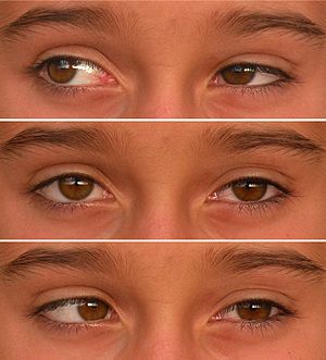
Duane syndrome
| Duane's syndrome | |
|---|---|
 |
|
| Duane Syndrome type I in left eye. 10-year-old girl. | |
| Classification and external resources | |
| Specialty | ophthalmology |
| ICD-10 | H50.8 |
| ICD-9-CM | 378.71 |
| OMIM | 126800 604356 |
| DiseasesDB | 30810 |
| eMedicine | oph/326 |
| MeSH | D004370 |
| GeneReviews | |
| Orphanet | 233 |
Duane syndrome is a congenital rare type of strabismus most commonly characterized by the inability of the eye to move outwards. The syndrome was first described by ophthalmologists Jakob Stilling (1887) and Siegmund Türk (1896), and subsequently named after Alexander Duane, who discussed the disorder in more detail in 1905.
Other names for this condition include: Duane's retraction syndrome, eye retraction syndrome, retraction syndrome, congenital retraction syndrome and Stilling-Türk-Duane Syndrome.
The characteristic features of the syndrome are:
While usually isolated to the eye abnormalities, Duane syndrome can be associated with other problems including cervical spine abnormalities Klippel-Feil syndrome, Goldenhar syndrome, heterochromia, and congenital deafness.
Duane syndrome is most probably a miswiring of the eye muscles, causing some eye muscles to contract when they shouldn't and other eye muscles not to contract when they should. Alexandrakis and Saunders found that in most cases the abducens nucleus and nerve are absent or hypoplastic, and the lateral rectus muscle is innervated by a branch of the oculomotor nerve. This view is supported by the earlier work of Hotchkiss et al. who reported on the autopsy findings of two patients with Duane's syndrome. In both cases the sixth cranial nerve nucleus and nerve was absent, and the lateral rectus muscle was innervated by the inferior division of the third or oculomotor nerve. This misdirection of nerve fibres results in opposing muscles being innervated by the same nerve. Thus, on attempted abduction, stimulation of the lateral rectus via the oculomotor nerve will be accompanied by stimulation of the opposing medial rectus via the same nerve; a muscle which works to adduct the eye. Thus, co-contraction of the muscles takes place, limiting the amount of movement achievable and also resulting in retraction of the eye into the socket. They also noticed mechanical factors and considered them secondary to loss of innervation: During corrective surgery fibrous attachments have been found connecting the horizontal recti and the orbital walls and fibrosis of the lateral rectus has been confirmed by biopsy. This fibrosis can result in the lateral rectus being 'tight' and acting as a tether or leash. Co-contraction of the medial and lateral recti allows the globe to slip up or down under the tight lateral rectus producing the up and down shoots characteristic of the condition.
...
Wikipedia
