Cerebellar vermis
| Cerebellar vermis | |
|---|---|

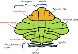
Schematic representation of the major anatomical subdivisions of the cerebellum. Superior view of an "unrolled" cerebellum, placing the vermis in one plane.
|
|

Under surface of the cerebellum. ("Tuber vermis" labeled at bottom.)
|
|
| Details | |
| Part of | Cerebellum |
| Identifiers | |
| Latin | vermis cerebelli |
| NeuroNames | ancil-146 |
| NeuroLex ID | Vermis |
| Dorlands /Elsevier |
v_06/12854118 |
| TA | A14.1.07.006 |
| FMA | 76928 |
|
Anatomical terms of neuroanatomy
[]
|
|
The cerebellar vermis (Latin for worm) is located in the medial, cortico-nuclear zone of the cerebellum, residing in the posterior fossa of the cranium. The primary fissure in the vermis curves ventrolaterally to the superior surface of the cerebellum, dividing it into anterior and posterior lobes. Functionally, the vermis is associated with bodily posture and locomotion. The vermis is included within the spinocerebellum and receives somatic sensory input from the head and proximal body parts via ascending spinal pathways.
The cerebellum develops in a rostro-caudal manner, with rostral regions in the midline giving rise to the vermis, and caudal regions developing into the cerebellar hemispheres. By 4 months of prenatal development, the vermis becomes fully foliated, while development of the hemispheres lags by 30–60 days. Postnatally, proliferation and organization of the cellular components of the cerebellum continues, with completion of the foliation pattern by 7 months of life and final migration, proliferation, and arborization of cerebellar neurons by 20 months.
...
Wikipedia
