Arbor vitae (anatomy)
| Arbor vitae | |
|---|---|

Figure shows Cerebellum and surrounding regions; sagittal view of one hemisphere. A: Midbrain. B: Pons. C: Medulla. D: Spinal cord. E: Fourth ventricle. F: Arbor vitae (in pink). G: Tonsil. H: Anterior lobe. I: Posterior lobe.
|
|

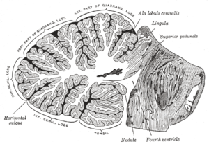
Sagittal section of the cerebellum, near the junction of the vermis with the hemisphere. ("arbor vitae" visible as white space to left, but not labelled.)
|
|
| Details | |
| Identifiers | |
| Latin | arbor vitae cerebelli |
| NeuroNames | hier-689 |
| NeuroLex ID | Arbor Vitae |
| Dorlands /Elsevier |
a_56/12149382 |
| TA | A14.1.07.401 |
| FMA | 72541 |
|
Anatomical terms of neuroanatomy
[]
|
|
The arbor vitae /ˌɑːrbɔːr ˈvaɪtiː/ (Latin for "tree of life") is the cerebellar white matter, so called for its branched, tree-like appearance. In some ways it more resembles a fern and is present in both cerebellar hemispheres. It brings sensory and motor information to and from the cerebellum. The arbor vitae is located deep in the cerebellum. Situated within the arbor vitae are the deep cerebellar nuclei; the dentate, globose, emboliform and the fastigial nuclei. These four different structures lead to the efferent projections of the cerebellum.
The arbor vitae is subject to pathologies such as a cerebellar hemorrhage. Cerebellar hemorrhages arise from tumors, trauma and arteriovenous malformations among other things. The cells in the arbor vitae could also be infected by pathogens which might cause lasting damage, this in turn could lead to cerebellar ataxia.
Godfrey Blount's 1899 book Arbor Vitae was ‘a book on the nature and development of imaginative design for the use of teachers and craftsmen’.
Arbor vitae
...
Wikipedia
