Cavernous hemangioma
| Cavernous hemangioma | |
|---|---|
 |
|
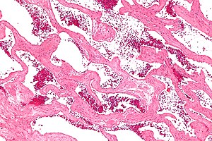
| Micrograph of a cavernous liver hemangioma. H&E stain. | |
| Classification and external resources |
Cavernous hemangioma, also called cavernous angioma, cavernoma, or cerebral cavernous malformation (CCM) (when referring to presence in the brain) is a type of blood vessel malformation or hemangioma, where a collection of dilated blood vessels form a benign tumor. Because of this malformation, blood flow through the cavities, or caverns, is slow. Additionally, the cells that form the vessels do not form the necessary junctions with surrounding cells. Also, the structural support from the smooth muscle is hindered, causing leakage into the surrounding tissue. It is the leakage of blood, known as a hemorrhage from these vessels that causes a variety of symptoms known to be associated with this disease.
Cavernous hemangiomas can arise nearly anywhere in the body where there are blood vessels and are considered to be benign tumours. They are often described as raspberry-like because of the bubble-like caverns. Unlike the capillary hemangiomas, cavernous ones can be disfiguring and do not tend to regress. Most cases of cavernomas are congenital, however they can develop over the course of a lifetime. While there is no definitive cause, research shows that genetic mutations result in the onset. Congenital hemangiomas that appear on the skin are known as either vascular or red birthmarks.
Gradient-Echo T2WI magnetic resonance imaging (MRI) is most sensitive method for diagnosing cavernous hemangiomas. MRI is such a powerful tool for diagnosis, it has led to an increase in diagnosis of cavernous hemangiomas since the technology's advent in the 1980s. The radiographic appearance is most commonly described as "popcorn" or "mulberry"-shaped.Computed tomography (CT) scanning is not a sensitive or specific method for diagnosing cavernous hemangiomas.Angiography is typically not necessary, unless it is required to rule out other diagnoses. Additionally, biopsies can be obtained from tumor tissue for examination under a microscope. It is essential to diagnose cavernous hemangioma because treatments for this benign tumor are less aggressive than that of cancerous tumors, such as angiosarcoma. However, since MRI appearance is practically pathognomonic, biopsy is rarely needed for verification.
...
Wikipedia
