Cardiac magnetic resonance imaging
| Cardiac magnetic resonance imaging | |
|---|---|

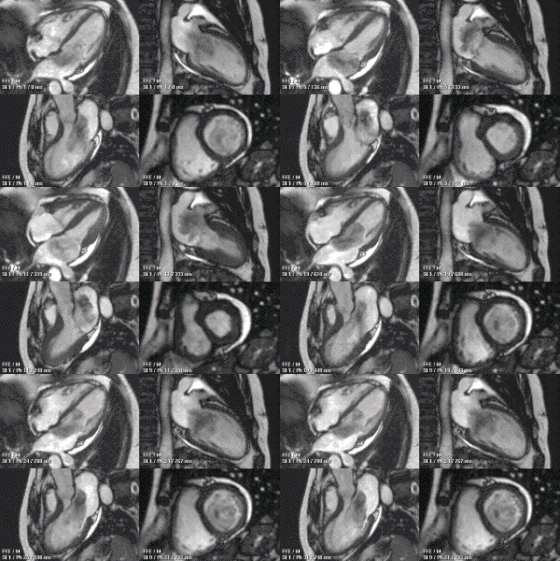
An example of CMR movies in different orientations of a cardiac tumor - in this case, an atrial myxoma.
|
|
| ICD-10-PCS | [1] |
| ICD-9-CM | 88.92 |
| OPS-301 code | 3-803, 3-824 |
Cardiovascular magnetic resonance imaging (CMR), sometimes known as cardiac MRI, is a medical imaging technology for the non-invasive assessment of the function and structure of the cardiovascular system. It is derived from and based on the same basic principles as magnetic resonance imaging (MRI) but with optimization for use in the cardiovascular system. These optimizations are principally in the use of ECG gating and rapid imaging techniques or sequences. By combining a variety of such techniques into protocols, key functional and morphological features of the cardiovascular system can be assessed.
In the investigation of cardiovascular disease the physician has a wide variety of tools available. The key disadvantages of CMR are limited availability, expense, and special skills/technical training needed to perform CMR (vs other types of MRI). New volumetric acquisitions can shorten and simplify the scan as they can replace several sequences, acquiring the entire cardiac volume at once. The key advantages are image quality, non-invasiveness, accuracy, versatility and no ionising radiation.
MRA (magnetic resonance angiography) can produce 3D and 4D images of blood vessels and the flow of blood through the vessels.
A good overview of the clinical indications for CMR can be found here and here
A good overview of the quantifiable results available from CMR may be found here.
There is no proven risk of biological harm from even very powerful static magnetic fields. However, genotoxic effects of cardiac MRI scanning have been demonstrated in vivo and in vitro, leading a recent review to recommend "a need for further studies and prudent use in order to avoid unnecessary examinations, according to the precautionary principle". In a comparison of genotoxic effects of MRI compared with those of CT scans, the cancer risk of MRI is unknown. As prior MRI risk research was only based on cell level experiments and there is no information on their relevance for developing malignant cells, these results do not provide definitive evidence for an actual cancer risk. This stands in contrast to the medical exposure of ionizing radiation which is clearly linked with cancer risk. Furthermore, dsDNA breaks as observed in these preliminary MRI studies are known to occur as a part of normal physiology including brain activity during wakefulness. Thus, MRI is still to be considered the safest of the advanced imaging techniques.
CMR uses the same basic principles as other MRI techniques with the addition of ECG gating. Most CMR uses only 1H nuclei MR, which are abundant in human tissue. By using magnetic fields and radiofrequency (RF) pulses, the patient's own 1H nuclei absorb and then emit energy, which can be measured and translated into images, without using ionising radiation.
...
Wikipedia
