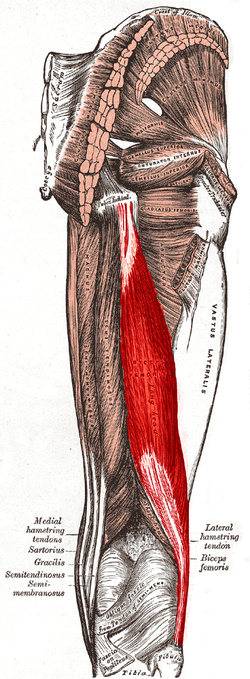
Biceps femoris
| Biceps Femoris | |
|---|---|

Posterior view of right leg. Long head of muscle highlighted in red, short head labeled in the lower part of the image
|
|
| Details | |
| Origin | tuberosity of the ischium, linea aspera, femur |
| Insertion | the head of the fibula which articulates with the back of the lateral tibial condyle |
| Artery | deep femoral artery, perforating arteries; long head of biceps femoris: perforating branches from profunda femoris artery |
| Nerve | long head: tibial nerve short head: common fibular nerve |
| Actions | flexes knee joint, laterally rotates knee joint (when knee is flexed), extends hip joint (long head only) |
| Antagonist | Quadriceps muscle |
| Identifiers | |
| Latin | musculus biceps femoris |
| Dorlands /Elsevier |
m_22/12548483 |
| TA | A04.7.02.032 |
| FMA | 22356 |
|
Anatomical terms of muscle
[]
|
|
The biceps femoris (/ˈbaɪsɛps ˈfɛmərᵻs/) is a muscle of the thigh located to the posterior, or back. As its name implies, it has two parts, one of which (the long head) forms part of the hamstrings muscle group.
It has two heads of origin;
The fibers of the long head form a belly, which passes obliquely downward and lateralward across the sciatic nerve to end in an aponeurosis which covers the posterior surface of the muscle, and receives the fibers of the short head; this aponeurosis becomes gradually contracted into a tendon, which is inserted into the lateral side of the head of the fibula, and by a small slip into the lateral condyle of the tibia.
At its insertion the tendon divides into two portions, which embrace the fibular collateral ligament of the knee-joint.
From the posterior border of the tendon a thin expansion is given off to the fascia of the leg. The tendon of insertion of this muscle forms the lateral hamstring; the common fibular (peroneal) nerve descends along its medial border.
The short head may be absent; additional heads may arise from the ischial tuberosity, the linea aspera, the medial supracondylar ridge of the femur, or from various other parts. The tendon of insertion may be attached to the Iliotibial band and to retinacular fibers of the lateral joint capsule.
...
Wikipedia
