Tibial nerve
| Tibial nerve | |
|---|---|

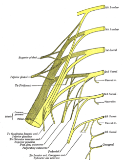
Plan of sacral and pudendal plexuses (Tibial nerve labelled at centre left)
|
|

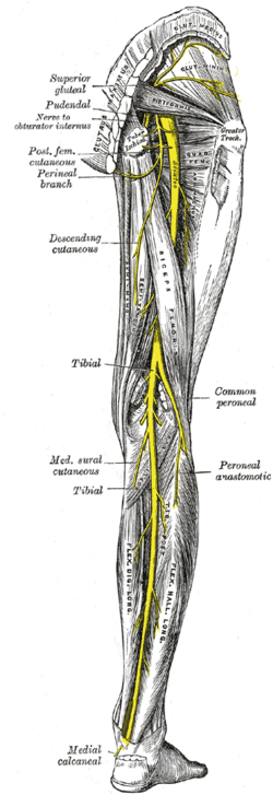
Nerves of the right lower extremity Posterior view.
|
|
| Details | |
| From | sacral plexus via sciatic nerve |
| To | medial plantar nerve, lateral plantar nerve |
| Innervates | Origin: flexor digitorum longus, flexor hallucis longus Medial: abductor hallucis, flexor digitorum brevis, flexor hallucis brevis, first lumbrical Lateral: quadratus plantae, flexor digiti minimi, adductor hallucis, the interossei, three lumbricals. and abductor digiti minimi |
| Identifiers | |
| Latin | Nervus tibialis |
| MeSH | A08.800.800.720.450.760.820 |
| TA | A14.2.07.058 |
| FMA | 19035 |
|
Anatomical terms of neuroanatomy
[]
|
|
The tibial nerve is a branch of the sciatic nerve. The tibial nerve passes through the popliteal fossa to pass below the arch of soleus.
In the popliteal fossa the nerve gives off branches to gastrocnemius, popliteus, soleus and plantaris muscles, an articular branch to the knee joint, and a cutaneous branch that will become the sural nerve. The sural nerve is joined by fibres from the common fibular nerve and runs down the calf to supply the lateral side of the foot.
Below the soleus muscle the nerve lies close to the tibia and supplies the tibialis posterior, the flexor digitorum longus and the flexor hallucis longus. The nerve passes into the foot running posterior to the medial malleolus. Here it is bound down by the flexor retinaculum in company with the posterior tibial artery.
In the foot, the nerve divides into medial and lateral plantar branches.
The medial plantar nerve supplies: the abductor hallucis, the flexor digitorum brevis, the flexor hallucis brevis and the first lumbrical. Cutaneous distribution of the medial plantar nerve is to the medial sole and medial three and one half toes, including the nail beds on the dorsum (like the median nerve in the hand).
...
Wikipedia
