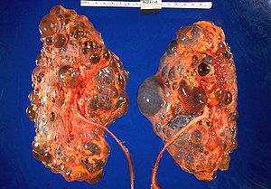
Autosomal dominant polycystic kidney disease
| Polycystic Kidney Disease | |
|---|---|
 |
|
| Polycystic kidneys | |
| Classification and external resources | |
| Specialty | medical genetics |
| ICD-10 | Q61 |
| ICD-9-CM | 753.1 |
| OMIM | 601313 173910 |
| DiseasesDB | 10262 |
| MedlinePlus | 000502 |
| eMedicine | radio/68 |
| MeSH | D016891 |
Autosomal dominant polycystic kidney disease (ADPKD, autosomal dominant PKD or adult-onset PKD) is the most prevalent, potentially lethal, monogenic human disorder. It is associated with large interfamilial and intrafamilial variability, which can be explained to a large extent by its genetic heterogeneity and modifier genes. It is also the most common of the inherited cystic kidney diseases — a group of disorders with related but distinct pathogenesis, characterized by the development of renal cysts and various extrarenal manifestations, which in case of ADPKD include cysts in other organs, such as the liver, seminal vesicles, pancreas, and arachnoid membrane, as well as other abnormalities, such as intracranial aneurysms and dolichoectasias, aortic root dilatation and aneurysms, mitral valve prolapse, and abdominal wall hernias. Over 50% of patients with ADPKD eventually develop end stage kidney disease and require dialysis or kidney transplantation. ADPKD is estimated to affect at least 1 in every 1000 individuals worldwide, making this disease the most common inherited kidney disorder with a diagnosed prevalence of 1:2000 and incidence of 1:3000-1:8000 in a global scale.
In many patients with ADPKD, kidney dysfunction is not clinically apparent until forty or fifty years of life. There is, however, an increasing body of evidence that suggests the formation of renal cysts starts in utero. Cysts initially form as small dilations in renal tubules, which then expand to form fluid-filled cavities of different sizes. Factors suggested to lead to cystogenesis include a germline mutation in one of the polycystin gene alleles, a somatic second hit that leads to the loss of the normal allele, and a third hit, which can be anything that triggers cell proliferation, leading to the dilation of the tubules. In the progression of the disease, continued dilation of the tubules through increased cell proliferation, fluid secretion, and separation from the parental tubule lead to the formation of cysts.
...
Wikipedia
