Atherogenesis
| Atherosclerosis | |
|---|---|
 |
|
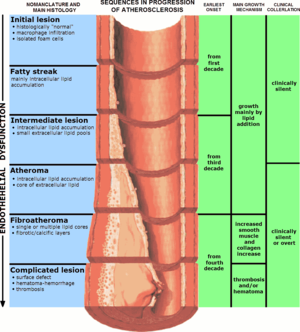
| The progression of atherosclerosis (narrowing exaggerated) | |
| Classification and external resources | |
| Specialty | Cardiology, angiology |
| ICD-10 | I25.0,I25.1,I70 |
| ICD-9-CM | 440, 414.0 |
| DiseasesDB | 1039 |
| MedlinePlus | 000171 |
| eMedicine | med/182 |
| Patient UK | Atherosclerosis |
| MeSH | D050197 |
Atherosclerosis (also known as arteriosclerotic vascular disease or ASVD) is a specific form of arteriosclerosis in which an artery-wall thickens as a result of invasion and accumulation of white blood cells (WBCs) (foam cell) and proliferation of intimal-smooth-muscle cell creating an atheromatous (fibrofatty) plaque.
The accumulation of the white blood cells is termed "fatty streaks" early on because of the appearance being similar to that of marbled steak. These accumulations contain both living, active WBCs (producing inflammation) and remnants of dead cells, including cholesterol and triglycerides. The remnants eventually include calcium and other crystallized materials within the outermost and oldest plaque. The "fatty streaks" reduce the elasticity of the artery walls. However, they do not affect blood flow for decades because the artery muscular wall enlarges at the locations of plaque. The wall stiffening may eventually increase pulse pressure; widened pulse pressure is one possible result of advanced disease within the major arteries.
Atherosclerosis is therefore a syndrome affecting arterial blood vessels due to a chronic inflammatory response of WBCs in the walls of arteries. This is promoted by low-density lipoproteins (LDL, plasma proteins that carry cholesterol and triglycerides) without adequate removal of fats and cholesterol from the macrophages by functional high-density lipoproteins (HDL). It is commonly referred to as a "hardening" or furring of the arteries. It is caused by the formation of multiple atheromatous plaques within the arteries.
The plaque is divided into three distinct components:
Atherosclerosis is a chronic disease that remains asymptomatic for decades. Atherosclerotic lesions, or atherosclerotic plaques, are separated into two broad categories: Stable and unstable (also called vulnerable). The pathobiology of atherosclerotic lesions is very complicated, but generally, stable atherosclerotic plaques, which tend to be asymptomatic, are rich in extracellular matrix and smooth muscle cells. On the other hand, unstable plaques are rich in macrophages and foam cells, and the extracellular matrix separating the lesion from the arterial lumen (also known as the fibrous cap) is usually weak and prone to rupture. Ruptures of the fibrous cap expose thrombogenic material, such as collagen, to the circulation and eventually induce thrombus formation in the lumen. Upon formation, intraluminal thrombi can occlude arteries outright (e.g., coronary occlusion), but more often they detach, move into the circulation, and eventually occlude smaller downstream branches causing thromboembolism. Apart from thromboembolism, chronically expanding atherosclerotic lesions can cause complete closure of the lumen. Chronically expanding lesions are often asymptomatic until lumen stenosis is so severe (usually over 80%) that blood supply to downstream tissue(s) is insufficient, resulting in ischemia.
...
Wikipedia
