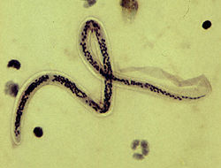
Wuchereria bancrofti
| Wuchereria bancrofti | |
|---|---|
 |
|
| Microfilar of W. bancrofti, from a patient seen in Haiti - the thick blood smears are stained with hematoxylin | |
| Scientific classification | |
| Kingdom: | Animalia |
| Phylum: | Nematoda |
| Class: | Secernentea |
| Order: | Spirurida |
| Suborder: | Spirurina |
| Family: | Onchocercidae |
| Genus: | Wuchereria |
| Species: | W. bancrofti |
| Binomial name | |
|
Wuchereria bancrofti (Cobbold, 1877) Seurat, 1921 |
|
| Wuchereria bancrofti | |
|---|---|
| Classification and external resources | |
| Specialty | infectious disease |
| ICD-10 | B74.0 |
| ICD-9-CM | 125.0 |
| DiseasesDB | 29234 |
Wuchereria bancrofti is a human parasitic roundworm that is the major cause of lymphatic filariasis. It is one of the three parasitic worms, together with Brugia malayi and B. timori, that infect the lymphatic system to cause lymphatic filariasis. These filarial worms are spread by a variety of mosquito vector species. W. bancrofti is the most prevalent of the three and affects over 120 million people, primarily in Central Africa and the Nile delta, South and Central America, the tropical regions of Asia including southern China, and the Pacific islands. If left untreated, the infection can develop into a chronic disease called elephantiasis. In rare conditions it also causes tropical eosinophilia, an asthmatic disease. Limited treatment modalities exist and no vaccines have been developed.
The effects of W. bancrofti were documented early in ancient text. Ancient Greek and Roman writers noted the similarities between the enlarged limbs and cracked skin of infected individuals to that of elephants. Since then, this condition has been commonly known as elephantiasis. However, this is a misnomer, since elephantiasis literally translates to “a disease caused by elephants”.
W. bancrofti was named after physician Otto Wucherer and parasitologist Joseph Bancroft, both of whom extensively studied filarial infections.
W. bancrofti is speculated to have been brought to the New World by the slave trade. Once it was introduced to the New World, this filarial worm disease persisted throughout the areas surrounding Charleston, South Carolina until its sudden disappearance in the 1920s.
As a dioecious worm, W. bancrofti exhibits sexual dimorphism. The adult worm is long, cylindrical, slender, and smooth with rounded ends. It is white in colour and almost transparent. The body is quite delicate making it difficult to remove from tissues. It has a short cephalic or head region connected to the main body by a short neck which appears as a constriction. There are dark spots which are dispersed nuclei throughout the body cavity, with no nuclei at the tail tip. Male and female can be differentiated by size and structure of tail tip. The male worm is smaller, 40 millimetres (1.6 in) long and 100 micrometres (0.0039 in) wide, and features a ventrally curved tail. The tip of the tail has 15 pairs of minute caudal papillae, the sensory organs. The anal region is an elaborate structure consisting of 12 pairs of papillae, of which 8 are in front and 4 are behind the anus. In contrast, the female is 60 millimetres (2.4 in) to 100 millimetres (3.9 in) long and 300 micrometres (0.012 in) wide, nearly three times larger in diameter than the male. Its tail gradually tapers and rounded at the tip. There are no additional sensory structures. Its vulva lies towards the anterior region, about 0.25 mm from the head. Adult male and female are most often coiled together and are difficult to separate. Females are ovoviviparous and can produce thousands of juveniles known as microfilariae.
...
Wikipedia
