Vitreous fluid
| Vitreous humour | |
|---|---|

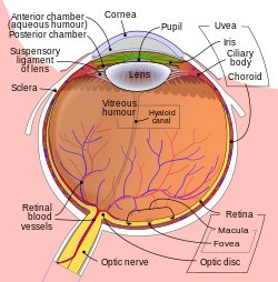
Schematic diagram of the human eye.
|
|
| Details | |
| Identifiers | |
| Latin | humor vitreus |
| TA |
A15.2.06.014 A15.2.06.008 |
| FMA | 67388 |
|
Anatomical terminology
[]
|
|
The vitreous body is the clear gel that fills the space between the lens and the retina of the eyeball of humans and other vertebrates. It is often referred to as the vitreous humour or simply "the vitreous".
The vitreous humour is a transparent, colorless, gelatinous mass that fills the space in the eye between the lens and the retina. It is present at birth. Produced by cells in the non-pigmented portion of the ciliary body, the vitreous humour is derived from embryonic mesenchyme cells, which degenerate after birth.
The vitreous humour is in contact with the retina and helps to keep it in place by pressing it against the choroid. It does not adhere to the retina, except at the optic nerve disc and the ora serrata (where the retina ends anteriorly), at the Wieger-band, the dorsal side of the lens. It is not connected at the macula, the area of the retina which provides finer detail and central vision.
Aquaporin-4 in Müller cell in rats, transports water to the vitreous body.
The vitreous has many anatomical landmarks, including the hyaloid membrane, Berger's space, space of Erggelet, Wieger's ligament, Cloquet's canal and the space of Martegiani.
Surface features:
Internal structures of the vitreous
Named tracts
The nature and composition of the vitreous humour changes over the course of life. At birth, the fluid is homogenous and finely striated (except Cloquet's canal). In adolescence, the vitreous cortex becomes more dense and vitreous tracts develop; and in adulthood, the tracts become better defined and sinuous. Central vitreous liquefies, fibrillar degeneration occurs, and the tracts break up (syneresis).
...
Wikipedia
