Visual area MT
| Visual cortex | |
|---|---|

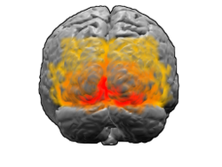
View of the brain from behind. Red = Brodmann area 17 (primary visual cortex); orange = area 18; yellow = area 19
|
|

Brain shown from the side, facing left. Above: view from outside, below: cut through the middle. Orange = Brodmann area 17 (primary visual cortex)
|
|
| Details | |
| Identifiers | |
| Latin | Cortex visualis |
| NeuroLex ID | Visual cortex primary |
| Dorlands /Elsevier |
c_57/12261838 |
| FMA | 242644 |
|
Anatomical terms of neuroanatomy
[]
|
|
The visual cortex of the brain is a part of the cerebral cortex that plays an important role in processing visual information. It is located in the occipital lobe in the back of the skull.
Visual information coming from the eye, goes through the lateral geniculate nucleus, that is located in the thalamus, and then reaches the visual cortex. The part of the visual cortex that receives the sensory inputs from the thalamus is the primary visual cortex, also known as Visual area one(V1), and the striate cortex. The extrastriate areas consist of visual areas two (V2), three (V3), four (V4), and five (V5).
Both hemispheres of the brain contain a visual cortex; the visual cortex in the left hemisphere receives signals from the right visual field, and the visual cortex in the right hemisphere receives signals from the left visual field.
The primary visual cortex (V1) is located in and around the calcarine fissure in the occipital lobe. Each hemisphere's V1 receives information directly from its ipsilateral lateral geniculate nucleus that receives signals from the contralateral visual hemifield.
Neurons in the visual cortex fire action potentials when visual stimuli appear within their receptive field. By definition, the receptive field is the region within the entire visual field that elicits an action potential. But, for any given neuron, it may respond best to a subset of stimuli within its receptive field. This property is called neuronal tuning. In the earlier visual areas, neurons have simpler tuning. For example, a neuron in V1 may fire to any vertical stimulus in its receptive field. In the higher visual areas, neurons have complex tuning. For example, in the inferior temporal cortex (IT), a neuron may fire only when a certain face appears in its receptive field.
...
Wikipedia
