Vesiculæ seminales
| Seminal vesicle | |
|---|---|

Human Male Anatomy
|
|

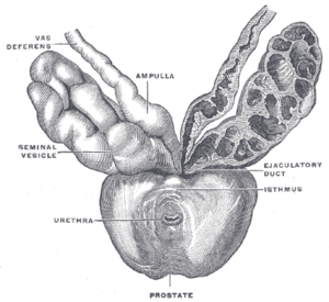
Prostate with seminal vesicles and seminal ducts, viewed from in front and above.
|
|
| Details | |
| Precursor | Wolffian duct |
| Artery | Inferior vesical artery, middle rectal artery |
| Lymph | External iliac lymph nodes, internal iliac lymph nodes |
| Identifiers | |
| Latin | Vesiculae seminales |
| MeSH | A05.360.444.713 |
| FMA | 19386 |
|
Anatomical terminology
[]
|
|
The seminal vesicles (Latin: glandulae vesiculosae), vesicular glands, or seminal glands, are a pair of simple tubular glands posteroinferior to the urinary bladder of some male mammals. Seminal vesicles are located within the pelvis. They secrete fluid that partly composes the semen.
The seminal vesicles are a pair of glands that are positioned below the urinary bladder and lateral to the vas deferens. Each vesicle consists of a single tube folded and coiled on itself, with occasional diverticula in its wall.
The excretory duct of each seminal gland unites with the corresponding vas deferens to form the two ejaculatory ducts, which immediately pass through the substance of the prostate gland before opening separately into the verumontanum of the prostatic urethra.
Each seminal vesicle spans approximately 5 cm, though its full unfolded length is approximately 10 cm, but it is curled up inside the gland's structure.
Each vesicle forms as an outpocketing of the wall of the ampulla of one vas deferens. The seminal vesicles develop as one of three structures of the male reproductive system that develops at the junction between the urethra and vas deferens. The vas deferens is derived from the mesonephric duct, a structure that develops from mesoderm.
Under microscopy, the seminal vesicles can be seen to have a mucosa, consisting of a lining of interspersed columnar cells and a lamina propria; and a thick muscular wall. The lumen of the glands is highly irregular and stores secretions from the glands of the vesicles. In detail:
...
Wikipedia
