Ventricle (brain)
| Ventricular system | |
|---|---|

The ventricular system accounts for the production and circulation of cerebrospinal fluid.
|
|

Rotating 3D rendering of the four ventricles and connections. From top to bottom:
Blue - Lateral ventricles Cyan - Interventricular foramina (Monro) Yellow - Third ventricle Red - Cerebral aqueduct (Sylvius) Purple - fourth ventricle Green - continuous with the central canal (Apertures to subarachnoid space are not visible) |
|
| Details | |
| Identifiers | |
| Latin | Ventriculi cerebri |
| MeSH | A08.186.211.276 |
| NeuroNames | ancil-192 |
| FMA | 78447 |
|
Anatomical terms of neuroanatomy
[]
|
|
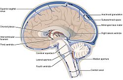
The ventricular system is a set of four interconnected cavities (ventricles) in the brain, where the cerebrospinal fluid (CSF) is produced. Within each ventricle is a region of choroid plexus, a network of ependymal cells involved in the production of CSF. The ventricular system is continuous with the central canal of the spinal cord (from the fourth ventricle) allowing for the flow of CSF to circulate. All of the ventricular system and the central canal of the spinal cord are lined with ependyma, a specialised form of epithelium.
The system comprises four ventricles:
There are several foramina, openings acting as channels, that connect the ventricles. The interventricular foramina (also called the foramina of Monro) connect the lateral ventricles to the third ventricle through which the cerebrospinal fluid can flow.
The four cavities of the human brain are called ventricles. The two largest are the lateral ventricles in the cerebrum; the third ventricle is in the diencephalon of the forebrain between the right and left thalamus; and the fourth ventricle is located at the back of the pons and upper half of the medulla oblongata of the hindbrain. The ventricles are concerned with the production and circulation of cerebrospinal fluid
The structures of the ventricular system are embryologically derived from the neural canal, the centre of the neural tube.
As the part of the primitive neural tube that will develop into the brainstem, the neural canal expands dorsally and laterally, creating the fourth ventricle, whereas the neural canal that does not expand and remains the same at the level of the midbrain superior to the fourth ventricle forms the cerebral aqueduct. The fourth ventricle narrows at the obex (in the caudal medulla), to become the central canal of the spinal cord.
...
Wikipedia
