Ventral nucleus of the lateral lemniscus
| Lateral lemniscus | |
|---|---|

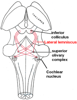
Lateral lemniscus in red, as it connects the cochlear nucleus, superior olivary nucleus and the inferior colliculus. Seen from behind.
|
|
| Details | |
| Identifiers | |
| Latin | lemniscus lateralis |
| NeuroNames | hier-605 |
| NeuroLex ID | Lateral lemiscus |
| Dorlands /Elsevier |
l_06/12483107 |
| TA |
A14.1.05.317 A14.1.08.670 A14.1.06.204 |
| FMA | 72502 |
|
Anatomical terms of neuroanatomy
[]
|
|
The lateral lemniscus is a tract of axons in the brainstem that carries information about sound from the cochlear nucleus to various brainstem nuclei and ultimately the contralateral inferior colliculus of the midbrain. Three distinct, primarily inhibitory, cellular groups are located interspersed within these fibers, and are thus named the nuclei of the lateral lemniscus.
The brainstem nuclei include:
Fibers leaving these brainstem nuclei ascending to the inferior colliculus rejoin the lateral lemniscus. In that sense, this is not a 'lemniscus' in the true sense of the word (second order, decussated sensory axons), as there is third (and out of the lateral superior olive, fourth) order information coming out of some of these brainstem nuclei.
The lateral lemniscus is located where the cochlear nuclei and the pontine reticular formation (PRF) crossover. The PRF descends the reticulospinal tract where it innervates motor neurons and spinal interneurons. It is the main auditory tract in the brainstem that connects the superior olivary complex (SOC) with the inferior colliculus (IC). The dorsal cochlear nucleus (DCN) has input from the LL and output to the contralateral LL via the ipsilateral and contralateral Dorsal Acoustic Stria.
There are three small nuclei on each of the lateral lemnisci: the ventral, dorsal, and the intermediate. The two lemnisci communicate via the commissural fibers of Probst.
The function of the lateral lemniscus is not known; however it has good temporal resolution compared to other cells higher than the cochlear nuclei and is sensitive to both timing and amplitude changes in sound. It is also involved in the acoustic startle reflex; the most likely region for this being the VNLL.
The cells of the DNLL respond best to bilateral inputs, and have onset and complexity tuned sustained responses. The nucleus is primarily GABAergic, and projects bilaterally to the inferior colliculus, and contralaterally to the DNLL, with different populations of cells projecting to each IC.
...
Wikipedia
