Uveoscleral tract
| Aqueous humor | |
|---|---|

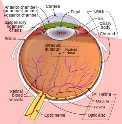
Schematic diagram of the human eye.
|
|
| Details | |
| Identifiers | |
| Latin | humor aquosus |
| TA | A15.2.06.002 |
| FMA | 58819 |
|
Anatomical terminology
[]
|
|
The aqueous humour is a transparent, watery fluid similar to plasma, but containing low protein concentrations. It is secreted from the ciliary epithelium, a structure supporting the lens. It fills both the anterior and the posterior chambers of the eye, and is not to be confused with the vitreous humour, which is located in the space between the lens and the retina, also known as the posterior cavity or vitreous chamber.
The main eye structures related to aqueous humour dynamics are the ciliary body (the site of aqueous humour production), and the trabecular meshwork and the uveoscleral pathway (the principal locations of aqueous humour outflow).
Aqueous humour is secreted into the posterior chamber by the ciliary body, specifically the non-pigmented epithelium of the ciliary body (pars plicata). It flows through the narrow cleft between the front of the lens and the back of the iris, to escape through the pupil into the anterior chamber, and then to drain out of the eye via the trabecular meshwork. From here, it drains into Schlemm's canal by one of two ways: directly, via aqueous vein to the episcleral vein, or indirectly, through collector channels to the episcleral vein by intrascleral plexus and eventually into the veins of the orbit. 5 alpha-dihydrocortisol, an enzyme inhibited by 5-alpha reductase inhibitors, may be involved in production of aqueous humour.
Aqueous humour is continually produced by the ciliary processes and this rate of production must be balanced by an equal rate of aqueous humour drainage. Small variations in the production or outflow of aqueous humour will have a large influence on the intraocular pressure.
The drainage route for aqueous humour flow is first through the posterior chamber, then the narrow space between the posterior iris and the anterior lens (contributes to small resistance), through the pupil to enter the anterior chamber. From there, the aqueous humour exits the eye through the trabecular meshwork into Schlemm's canal (a channel at the limbus, i.e., the joining point of the cornea and sclera, which encircles the cornea) It flows through 25–30 collector canals into the episcleral veins. The greatest resistance to aqueous flow is provided by the trabecular meshwork (esp. the juxtacanalicular part), and this is where most of the aqueous outflow occurs. The internal wall of the canal is very delicate and allows the fluid to filter due to high pressure of the fluid within the eye. The secondary route is the uveoscleral drainage, and is independent of the intraocular pressure, the aqueous flows through here, but to a lesser extent than through the trabecular meshwork (approx. 10% of the total drainage whereas by trabecular meshwork 90% of the total drainage).
...
Wikipedia
