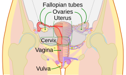
Uterine tubes
| Fallopian tubes | |
|---|---|

Schematic frontal view of female anatomy
|
|

Vessels of the uterus and its appendages, rear view. (Fallopian tubes visible at top right and top left.)
|
|
| Details | |
| Precursor | Müllerian duct |
| Artery | tubal branches of ovarian artery, tubal branch of uterine artery via mesosalpinx |
| Lymph | lumbar lymph nodes |
| Identifiers | |
| Latin | Tuba uterina |
| Greek | Salpinx |
| MeSH | A05.360.319.114.373 |
| Dorlands /Elsevier |
12827008 |
| TA | A09.1.02.001 |
| FMA | 18245 |
|
Anatomical terminology
[]
|
|
The Fallopian tubes, also known as uterine tubes or salpinges (singular salpinx), are two very fine tubes lined with ciliated epithelia, leading from the ovaries of female mammals into the uterus, via the uterotubal junction. In non-mammalian vertebrates, the equivalent structures are called oviducts.
In a woman's body the tube allows passage of the egg from the ovary to the uterus. Its different segments are (lateral to medial): the infundibulum with its associated fimbriae near the ovary, the ampullary region that represents the major portion of the lateral tube, the isthmus which is the narrower part of the tube that links to the uterus, and the interstitial (also known as intramural) part that transverses the uterine musculature. The ostium is the point where the tubal canal meets the peritoneal cavity, while the uterine opening of the Fallopian tube is the entrance into the uterine cavity, the uterotubal junction.
A cross section of Fallopian tube shows four distinct layers: serosa, subserosa, lamina propria and innermost mucosal layer. The serosa is derived from visceral peritoneum. Subserosa is composed of loose adventitious tissue, blood vessels, lymphatics, an outer longitudinal and inner circular smooth muscle coats. This layer is responsible for the peristaltic action of the Fallopian tubes. Lamina propria is a vascular connective tissue. There are two types of cells within the simple columnar epithelium of the Fallopian tube (oviduct). Ciliated cells predominate throughout the tube, but are most numerous in the infundibulum and ampulla. Estrogen increases the production of cilia on these cells. Interspersed between the ciliated cells are peg cells, which contain apical granules and produce the tubular fluid. This fluid contains nutrients for spermatozoa, oocytes, and zygotes. The secretions also promote capacitation of the sperm by removing glycoproteins and other molecules from the plasma membrane of the sperm. Progesterone increases the number of peg cells, while estrogen increases their height and secretory activity. Tubal fluid flows against the action of the ciliae, that is toward the fimbrial end.
...
Wikipedia
