Trochlear process
| Calcaneus | |
|---|---|

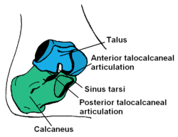
The calcaneus forms the bony part of the heel. It forms a joint with the talus bone, the subtalar joint.
|
|

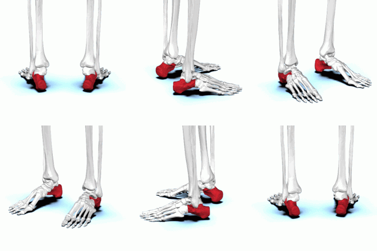
Bones of the foot, with the calcaneus shown in red
|
|
| Details | |
| Identifiers | |
| Latin | Calcaneus, Calcaneum, Os calcis |
| MeSH | A02.835.232.043.300.710.300 |
| TA | A02.5.11.001 |
| FMA | 24496 |
|
Anatomical terms of bone
[]
|
|
In humans, the calcaneus (/kælˈkeɪniːəs/; from the Latin calcaneus or calcaneum, meaning heel) or heel bone is a bone of the tarsus of the foot which constitutes the heel. In some other animals, it is the point of the hock.
In humans, the calcaneus is the largest of the tarsal bones and the largest bone of the foot. The talus bone, calcaneus, and navicular bone are considered the proximal row of tarsal bones. In the calcaneus, several important structures can be distinguished:
The half of the bone closest to the heel is the calcaneal tubercle. On its lower edge on either side are its lateral and medial processes (serving as the origins of the abductor hallucis and abductor digiti minimi). The Achilles tendon is inserted into a roughened area on its superior side, the cuboid bone articulates with its anterior side, and on its superior side are three articular surfaces for the articulation with the talus bone. Between these superior articulations and the equivalents on the talus is the tarsal sinus (a canal occupied by the interosseous talocalcaneal ligament). At the upper and forepart of the medial surface of the calcaneus, below the middle talar facet, there is a horizontal eminence, the talar shelf (also sustentaculum tali), which gives attachment to the plantar calcaneonavicular (spring) ligament, tibiocalcaneal ligament, and medial talocalcaneal ligament. This eminence is concave above, and articulates with the middle calcaneal articular surface of the talus; below, it is grooved for the tendon of the flexor hallucis longus; its anterior margin gives attachment to the plantar calcaneonavicular ligament, and its medial margin to a part of the deltoid ligament of the ankle-joint.
...
Wikipedia
