Tractography
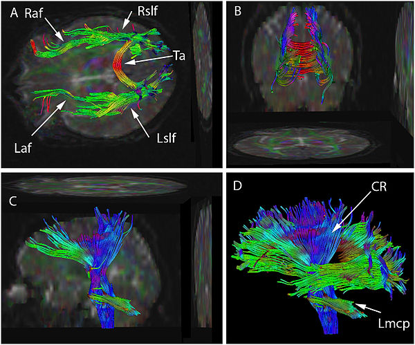
In neuroscience, tractography is a 3D modeling technique used to visually represent neural tracts using data collected by Difusion Weighted images (DWI). It uses special techniques of magnetic resonance imaging (MRI), and computer-based image analysis. The results are presented in two- and three-dimensional images.
In addition to the long tracts that connect the brain to the rest of the body, there are complicated neural networks formed by short connections among different cortical and subcortical regions. The existence of these bundles has been revealed by and biological techniques on post-mortem specimens. Brain tracts are not identifiable by direct exam, CT, or MRI scans. This difficulty explains the paucity of their description in neuroanatomy atlases and the poor understanding of their functions.
The MRI sequences used look at the symmetry of brain water diffusion. Bundles of fiber tracts make the water diffuse asymmetrically in a tensor, the major axis parallel to the direction of the fibers. The asymmetry here is called anisotropy. There is a direct relationship between the number of fibers and the degree of anisotropy.
...
Wikipedia

