Time-lapse microscopy

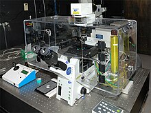
A time-lapse microscope. Notice the transparent cell incubator which is necessary to keep cells alive during observation.
|
|
| Other names | (Time-lapse) microcinematograph, (Time-lapse) video microscope, Time-lapse cinemicrograph |
|---|---|
| Uses | Observation of slow microscopic processes |
| Inventor | Jean Comandon and other contemporaries |
| Related items | Time-lapse photography, Live cell imaging |
Time-lapse microscopy is time-lapse photography applied to microscopy. Microscope image sequences are recorded and then viewed at a greater speed to give an accelerated view of the microscopic process.
Before the introduction of the video tape recorder in the 1960s, time-lapse microscopy recordings were made on photographic film. During this period, time-lapse microscopy was referred to as microcinematography. With the increasing use of video recorders, the term time-lapse video microscopy was gradually adopted. Today, the term video is increasingly dropped, reflecting that a digital still camera is used to record the individual image frames, instead of a video recorder.
Time-lapse microscopy can be used to observe any microscopic object over time. However, its main use is within cell biology to observe artificially cultured cells. Depending on the cell culture, different microscopy techniques can be applied to enhance characteristics of the cells as most cells are transparent.
To enhance observations further, cells have therefore traditionally been stained before observation. Unfortunately, the staining process kills the cells. The development of less destructive staining methods and methods to observe unstained cells has led to that cell biologists increasingly observe living cells. This is known as live cell imaging.
Time-lapse microscopy is the method that extends live cell imaging from a single observation in time to the observation of cellular dynamics over long periods of time. Time-lapse microscopy is primarily used in research, but is clinically used in IVF clinics as studies has proven it to increase pregnancy rates, lower abortion rates and predict aneuploidy
Modern approaches are further extending time-lapse microscopy observations beyond making movies of cellular dynamics. Traditionally, cells have been observed in a microscope and measured in a cytometer. Increasingly this boundary is blurred as cytometric techniques are being integrated with imaging techniques for monitoring and measuring dynamic activities of cells and subcellular structures.
...
Wikipedia
