Thoracic wall
| Thoracic wall | |
|---|---|

Body cavities
|
|

A transverse section of the thorax, showing the contents of the middle and the posterior mediastinum.
|
|
|
Anatomical terminology
[]
|
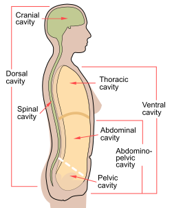
The thoracic wall or chest wall is the boundary of the thoracic cavity.
The bony skeletal part of the thoracic wall is the rib cage, and the rest is made up of muscle, skin, and fasciae.
The chest wall has 10 layers, namely skin, superficial fascia, deep fascia, serratus anterior, layer for ribs (containing intercostal muscles), and endothoracic fascia from superficial to deep. However, the muscular layers vary according to the region of the chest wall. For example, they may include muscles like pectoralis major or latissimus dorsi.
When not breathing for long and dangerous periods of time in cold water, a person's body undergoes great temporary changes to try to prevent death. It achieves this through the activation of the mammalian diving reflex, which has 3 main properties. Other than Bradycardia and Peripheral vasoconstriction, there is a blood shift which occurs only during very deep dives that affects the thoracic cavity (a chamber of the body protected by the thoracic wall.) When this happens, organ and circulatory walls allow plasma/water to pass freely throughout the thoracic cavity, so its pressure stays constant and the organs aren't crushed. In this stage, the lungs' alveoli fill up with blood plasma, which is reabsorbed when the organism leaves the pressurized environment. This stage of the diving reflex has been observed in humans (such as world champion freediver Martin Štěpánek) during extremely deep (over 90 metres or 300 ft) free dives.
In rare cases intentional or accidental trauma may lead to chest wall (thoracic wall) necrosis.
...
Wikipedia
