Thoracic vertebra 12
| thoracic vertebrae | |
|---|---|

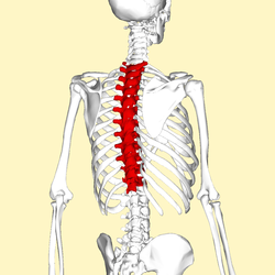
Position of the thoracic vertebrae (shown in red). In human, thoracic vertebrae consists of 12 bones. From top to down, T1, T2, …, T12.
|
|

A typical thoracic vertebra, seen from lateral side.
|
|
| Details | |
| Identifiers | |
| Latin | vertebrae thoracicae |
| MeSH | A02.835.232.834.892 |
| TA | A02.2.03.001 |
| FMA | 9139 72064, 9139 |
|
Anatomical terms of bone
[]
|
|
In vertebrates, thoracic vertebrae compose the middle segment of the vertebral column, between the cervical vertebrae and the lumbar vertebrae. In humans, there are twelve thoracic vertebrae and they are intermediate in size between the cervical and lumbar vertebrae; they increase in size going towards the lumbar vertebrae, with the lower ones being a lot larger than the upper. They are distinguished by the presence of facets on the sides of the bodies for articulation with the heads of the ribs, as well as facets on the transverse processes of all, except the eleventh and twelfth, for articulation with the tubercles of the ribs. By convention, the human thoracic vertebrae are numbered T1–T12, with the first one (T1) located closest to the skull and the others going down the spine toward the lumbar region.
These are the general characteristics of the second through eighth thoracic vertebrae. The first and ninth through twelfth vertebrae contain certain peculiarities, and are detailed below.
The bodies in the middle of the thoracic region are heart-shaped, and as broad in the antero-posterior as in the transverse direction. At the ends of the thoracic region they resemble respectively those of the cervical and lumbar vertebrae. They are slightly thicker behind than in front, flat above and below, convex from side to side in front, deeply concave behind, and slightly constricted laterally and in front. They present, on either side, two costal demi-facets, one above, near the root of the pedicle, the other below, in front of the inferior vertebral notch; these are covered with cartilage in the fresh state, and, when the vertebrae are articulated with one another, form, with the intervening intervertebral fibrocartilages, oval surfaces for the reception of the heads of the ribs.
The pedicles are directed backward and slightly upward, and the inferior vertebral notches are of large size, and deeper than in any other region of the vertebral column.
...
Wikipedia
