Thick ascending limb of loop of Henle
| Ascending limb of loop of Henle | |
|---|---|

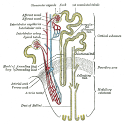
Scheme of renal tubule and its vascular supply. (Labeled at center left.)
|
|

Nephron ion flow diagram
|
|
| Details | |
| Identifiers | |
| Latin | tubulus rectus distalis, pars recta tubuli distalis |
| Dorlands /Elsevier |
t_22/12830078 |
| FMA | 17717 |
|
Anatomical terminology
[]
|
|
Within the nephron of the kidney, the ascending limb of the loop of Henle is a segment of the loop of Henle downstream of the descending limb, after the sharp bend of the loop. This part of the renal tubule is divided into a thin and thick ascending limb; the thick portion is also known as the distal straight tubule, in contrast with the distal convoluted tubule downstream.
The ascending limb of the loop of Henle is a direct continuation from the descending limb of loop of Henle, and one of the structures in the nephron of the kidney. The ascending limb has a thin and a thick segment. The ascending limb drains urine into the distal convoluted tubule.
The thin ascending limb is found in the medulla of the kidney, and the thick ascending limb can be divided into a part that is in the renal medulla and a part that is in the renal cortex. The ascending limb is much thicker than the descending limb.
As in the descending limb, the epithelium is simple squamous epithelium.
The thin ascending limb is impermeable to water; but is permeable to ions that cross by diffusion. Water moves out of the tubule and into the interstitium due to osmotic pressure created by the countercurrent system.
Functionally, the parts of the ascending limb in the medulla and cortex are very similar.
The medullary ascending limb is impermeable to water. Sodium (Na+), potassium (K+) and chloride (Cl−) ions are reabsorbed by active transport. K+ is passively transported along its concentration gradient through a K+ leak channel in the apical aspect of the cells, back into the lumen of the ascending limb. This K+ "leak" generates a positive electrochemical potential difference in the lumen. This drives more paracellular reabsorption of Na+, as well as other cations such as magnesium (Mg2+) and importantly calcium Ca2+ due to charge repulsion.
...
Wikipedia
