Tendinous arch of levator ani
| Levator ani | |
|---|---|

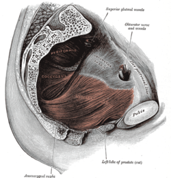
Left Levator ani seen from within.
|
|

Coronal section through the male anal canal. B. Cavity of urinary bladder V.D. Vas deferens. S.V. Seminal vesicle. R. Second part of rectum. A.C. Anal canal. L.A. Levator ani. I.S. Internal anal sphincter. E.S External anal sphincter.
|
|
| Details | |
| Origin | Inner surface of the side of the lesser pelvis |
| Insertion | Inner surface of coccyx, levator ani of opposite side, and into structures that penetrate it. |
| Artery | Inferior gluteal artery |
| Nerve | |
| Actions | Supports the viscera in pelvic cavity |
| Identifiers | |
| Latin | Musculus levator ani |
| TA | A04.5.04.002 |
| FMA | 19087 |
|
Anatomical terms of muscle
[]
|
|
pubococcygeus and iliococcygeus:
The levator ani is a broad, thin muscle, situated on either side of the pelvis. It is formed from three muscle components: the puborectalis, the pubococcygeus muscle (which includes the puborectalis) and the iliococcygeus muscle.
It is attached to the inner surface of each side of the lesser pelvis, and these unite to form the greater part of the pelvic floor. The coccygeus muscle completes the pelvic floor which is also called the pelvic diaphragm.
It supports the viscera in the pelvic cavity, and surrounds the various structures that pass through it.
The levator ani is the main pelvic floor muscle and contracts rhythmically during orgasm.
The levator ani is divided into three parts:
The iliococcygeus arises from the inner side of the ischium (the lower and back part of the hip bone) and from the posterior part of the tendinous arch of the obturator fascia, and is attached to the coccyx and anococcygeal body; it is usually thin, and may be absent, or be largely replaced by fibrous tissue. An accessory slip at its posterior part is sometimes named the iliosacralis.
The pubococcygeus muscle is then divided into three parts on its own.
The levator ani arises, in front, from the posterior surface of the superior pubic ramus lateral to the symphysis; behind, from the inner surface of the spine of the ischium; and between these two points, from the obturator fascia.
...
Wikipedia
