Temporomandibular
| Temporomandibular joint | |
|---|---|

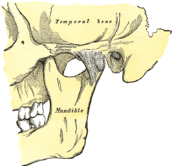
Articulation of the mandible. Lateral aspect.
|
|

Articulation of the mandible. Medial aspect.
|
|
| Details | |
| Artery | Superficial temporal artery |
| Nerve | Auriculotemporal nerve, masseteric nerve |
| Identifiers | |
| Latin | Articulatio temporomandibularis |
| Acronym(s) | TMJ |
| MeSH | A02.835.583.861 |
| Dorlands /Elsevier |
12161641 |
| TA | A03.1.07.001 |
| FMA | 54832 |
|
Anatomical terminology
[]
|
|
The temporomandibular joints (TMJ) are the two joints connecting the jawbone to the skull. It is a bilateral synovial articulation between the temporal bone of the skull above and the mandible below; it is from these bones that its name is derived.
The main components are the joint capsule, articular disc, mandibular condyles, articular surface of the temporal bone, temporomandibular ligament, stylomandibular ligament, sphenomandibular ligament, and lateral pterygoid muscle.
The capsule is a dense fibrous membrane that surrounds the joint and incorporates the articular eminence. It attaches to the articular eminence, the articular disc and the neck of the mandibular condyle.
The unique feature of the temporomandibular joint is the articular disc. The disc is composed of dense fibrous connective tissue that is positioned between the two bones that form the joint. The temporomandibular joints are one of the few synovial joints in the human body with an articular disc, another being the sternoclavicular joint. The disc divides each joint into two.These two compartments are synovial cavities, which consists of an upper and a lower synovial cavity. The synovial membrane lining the joint capsule produces the synovial fluid that fills these cavities.
The central area of the disc is avascular and lacks innervation, and, in contrast, the peripheral region has both blood vessels and nerves. Few cells are present, but fibroblasts and white blood cells are among these. The central area is also thinner but of denser consistency than the peripheral region, which is thicker but has a more cushioned consistency. The synovial fluid in the synovial cavities provides the nutrition for the avascular central area of the disc. With age, the entire disc thins and may undergo addition of cartilage in the central part, changes that may lead to impaired movement of the joint.
The lower joint compartment formed by the mandible and the articular disc is involved in rotational movement—this is the initial movement of the jaw when the mouth opens. The upper joint compartment formed by the articular disc and the temporal bone is involved in translational movement—this is the secondary gliding motion of the jaw as it is opened widely. The part of the mandible which mates to the under-surface of the disc is the condyle and the part of the temporal bone which mates to the upper surface of the disk is the articular fossa or glenoid fossa or mandibular fossa.
...
Wikipedia
