T-tubule
| T-tubule | |
|---|---|

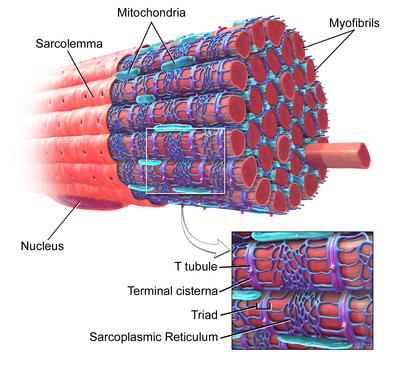
Figure 1: Skeletal muscle, with T-tubule labeled in zoomed in image.
|
|

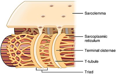
Narrow T-tubules permit the conduction of electrical impulses. The SR functions to regulate intracellular levels of calcium. Two terminal cisternae (where enlarged SR connects to the T-tubule) and one T-tubule comprise a triad—a “threesome” of membranes, with those of SR on two sides and the T-tubule sandwiched in-between them.
|
|
| Details | |
| Identifiers | |
| Latin | tubulus transversus |
| Code |
TH H2.00.05.2.01018 TH H2.00.05.2.02013 |
|
Anatomical terminology
[]
|
|
Transverse tubules (T-tubules) are extensions of the sarcolemma (muscle cell membrane) that penetrate into the centre of skeletal and cardiac muscle cells. T-tubules are highly specialised structures, that form within the first few weeks of life, containing large amounts of specific proteins known as ion channels that allow for electrical impulses (action potentials) travelling along the sarcolemma, to enter rapidly into the cell, to initiate muscle contraction.
Each muscle cell is surrounded by a sarcolemma. As T-tubules are simply continuations of the sarcolemma, their membranes are very similar to that of the cell membrane, consisting of two layers of lipid (fat) molecules (known as a lipid bilayer) with proteins, including: L-type calcium channels, sodium-calcium exchangers, calcium ATPases and Beta adrenoceptors, (see below) embedded within.
Despite what their name suggests, transverse-tubules are actually networks of tubules with both transverse (running at a right angle to the sarcolemma) sections, connected by axial/longitudinal tubules. Therefore, they are also known as the transverse-axial tubular system and they run alongside myofilament bundles (proteins that are responsible for muscle contraction).
The shape of the T-tubule system is produced and maintained by a variety of proteins. For example, a protein called Amphyphysin 2 is responsible to forming the structure of the T-tubule as well as ensuring that the appropriate proteins (in particular the L-type calcium channels) are located within the T-tubule membrane. Another example, is junctophilin 2. This protein helps to form a junction between the t-tubule membrane and an intracellular calcium store known as the sarcoplasmic reticulum (SR). This junction is vital for excitation-contraction coupling within muscle cells (see below). A final example is Tcap. Tcap helps with t-tubule development. There has been shown to be increased amounts of Tcap present in response to muscles stretching. This protein is therefore responsible for an increase in the number of t-tubules as muscles grow.
Skeletal and cardiac muscles are known collectively as striated muscles. This is because of their stripey appearance due to the contractile proteins (also known as myofilaments; in paricular actin and myosin) within them.
In cardiac muscle cells, T-tubules are between 20 and 450 nanometers in diameter (nm; 1 nm=0.000000001m) and are usually located in regions called Z-discs (this is where the actin fiilaments (mentioned above) anchor, within the cell). T-tubules within the heart are closely associated with a region of the sarcoplasmic reticulum known as terminal cisternae. This is known as a dyad.
...
Wikipedia
