Suspensory muscle of the duodenum
| Suspensory muscle of duodenum | |
|---|---|

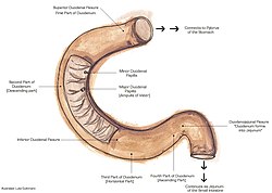
The duodenum. The suspensory muscle of the duodenum attaches to the duodenojejunal flexure, shown.
|
|

|
|
| Details | |
| System | Gastrointestinal |
| Origin | Connective tissue surrounding coeliac artery and superior mesenteric artery |
| Insertion | Third and fourth-parts of duodenum, duodenojejunal flexure |
| Nerve | Coeliac plexus, Superior mesenteric plexus |
| Actions | Facilitates movement of food; embryological role in fixating jejunum during gut rotation |
| Identifiers | |
| Latin | Musculus suspensorius duodeni, ligamentum suspensorium duodeni |
| Dorlands /Elsevier |
m_22/12551047 |
| TA | A05.6.02.011 |
| FMA | 20509 |
|
Anatomical terms of muscle
[]
|
|
The suspensory muscle of duodenum is a thin muscle connecting the junction between the duodenum, jejunum, and duodenojejunal flexure to connective tissue surrounding the superior mesenteric artery and coeliac artery. It is also known as the ligament of Treitz. The suspensory muscle most often connects to both the third and fourth parts of the duodenum, as well as the duodenojejunal flexure, although the attachment is quite variable.
The suspensory muscle marks the formal division between the first and second parts of the small intestine, the duodenum and the jejunum. This division is used to mark the difference between the upper and lower gastrointestinal tracts, which is relevant in clinical medicine as it may determine the source of bleeding in the gastrointestinal tract.
The suspensory muscle is derived from mesoderm and plays a role in the embryological rotation of the gut, by offering a point of fixation for the rotating gut. It is also thought to help digestion by widening the angle of the duodenojejunal flexure. Superior mesenteric artery syndrome is a rare abnormality caused by a congenitally-short suspensory muscle.
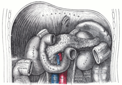
The duodenum and the jejunum are the first and second parts of the small intestine, respectively. The suspensory muscle of the duodenum marks their formal division. The suspensory muscle arises from the right crus of the diaphragm as it passes around the esophagus, continues as connective tissue around the stems of the celiac trunk (celiac artery) and superior mesenteric artery, passes behind the pancreas, and enters the upper part of the mesentery, inserting into the junction between the duodenum and jejunum, the duodenojejunal flexure. Here, the muscles are continuous with the muscular layers of the duodenum.
...
Wikipedia
