Supinator
| Supinator muscle | |
|---|---|

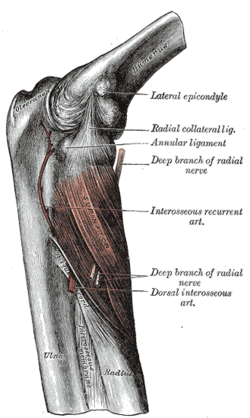
Posterior view of the supinator. (Right arm.)
|
|
| Details | |
| Origin | Lateral epicondyle of humerus, supinator crest of ulna, radial collateral ligament, annular ligament |
| Insertion | Lateral proximal radial shaft |
| Artery | Radial recurrent artery |
| Nerve | Deep branch of the radial nerve |
| Actions | Supinates forearm |
| Antagonist | Pronator teres, pronator quadratus |
| Identifiers | |
| Latin | musculus supinator |
| Dorlands /Elsevier |
m_22/12551025 |
| TA | A04.6.02.048 |
| FMA | 38512 |
|
Anatomical terms of muscle
[]
|
|
In human anatomy, the supinator is a broad muscle in the posterior compartment of the forearm, curved around the upper third of the radius. Its function is to supinate the forearm.
Supinator consists of two planes of fibers, between which the deep branch of the radial nerve ls. The two planes arise in common — the superficial one by tendinous (the initial portion of the muscle is actually just tendon) and the deeper by muscular fibers — from the supinator crest of ulna, the lateral epicondyle of humerus, the radial collateral ligament, and the annular radial ligament.
The superficial fibers (pars superficialis) surround the upper part of the radius, and are inserted into the lateral edge of the radial tuberosity and the oblique line of the radius, as low down as the insertion of the pronator teres. The upper fibers (pars profunda) of the deeper plane form a sling-like fasciculus, which encircles the neck of the radius above the tuberosity and is attached to the back part of its medial surface; the greater part of this portion of the muscle is inserted into the dorsal and lateral surfaces of the body of the radius, midway between the oblique line and the head of the bone.
The proximal aspect of the superficial head is known as the arcade of Frohse or the supinator arch.
It is innervated by the deep branch of the radial nerve. The deep branch then becomes the posterior interosseous nerve upon exiting the supinator muscle
...
Wikipedia
