Superior cluneal nerves
| Superior cluneal nerves | |
|---|---|

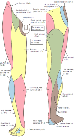
Cutaneous nerves of the right lower extremity. Front and posterior views. (Posterior division of lumbar visible in yellow at top right.)
|
|
| Details | |
| From | posterior branches of the lumbar nerves |
| Innervates | |
| Identifiers | |
| Latin | nervi clunium superiores |
| Dorlands /Elsevier |
n_0/12565406 |
| TA | A14.2.05.006 |
| FMA | 75468 |
|
Anatomical terms of neuroanatomy
[]
|
|
The superior cluneal nerves innervate the skin of the upper part of the . They are the terminal ends of lateral rami of the posterior rami of lumbar spinal nerves (L1, 2, 3).
Superior Cluneal Nerve Entrapment
The medial branch of the superior cluneal nerve passes over the iliac crest through a tunnel formed by the thoracolumbar fascia and the superior rim of the iliac crest. This branch of the superior cluneal nerve may become restricted in its osteofibrous tunnel against the iliac crest, just as osteofibrous tunnels affect other nerves, such as in carpal tunnel syndrome. The clinical symptoms include pain at low back which may radiate to the ipsilateral leg. The clinical signs include marked tenderness at iliac crest rim just above the dimple at the buttock and decreased touch sensation of the buttock just below the iliac crest. The treatment includes elimination of inappropriate use such as forward bending or acute twisting of the low back, NSAID therapy and local steroid injection. Surgical treatment by nerve decompression is used for cases of severe pain with failure of conservative treatment.
Diagram of the distribution of the cutaneous branches of the posterior divisions of the spinal nerves.
Areas of distribution of the cutaneous branches of the posterior divisions of the spinal nerves.
...
Wikipedia
